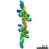
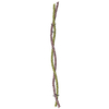
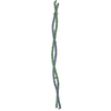
Journal: Nat Commun / Year: 2021 Title: Allostery through DNA drives phenotype switching. Authors: Gabriel Rosenblum / Nadav Elad / Haim Rozenberg / Felix Wiggers / Jakub Jungwirth / Hagen Hofmann / Abstract: Allostery is a pervasive principle to regulate protein function. Growing evidence suggests that also DNA is capable of transmitting allosteric signals. Yet, whether and how DNA-mediated allostery ...Allostery is a pervasive principle to regulate protein function. Growing evidence suggests that also DNA is capable of transmitting allosteric signals. Yet, whether and how DNA-mediated allostery plays a regulatory role in gene expression remained unclear. Here, we show that DNA indeed transmits allosteric signals over long distances to boost the binding cooperativity of transcription factors. Phenotype switching in Bacillus subtilis requires an all-or-none promoter binding of multiple ComK proteins. We use single-molecule FRET to demonstrate that ComK-binding at one promoter site increases affinity at a distant site. Cryo-EM structures of the complex between ComK and its promoter demonstrate that this coupling is due to mechanical forces that alter DNA curvature. Modifications of the spacer between sites tune cooperativity and show how to control allostery, which allows a fine-tuning of the dynamic properties of genetic circuits.
In the structure databanks used in Yorodumi, some data are registered as the other names, "COVID-19 virus" and "2019-nCoV". Here are the details of the virus and the list of structure data.
Jan 31, 2019. EMDB accession codes are about to change! (news from PDBe EMDB page)
EMDB accession codes are about to change! (news from PDBe EMDB page)
The allocation of 4 digits for EMDB accession codes will soon come to an end. Whilst these codes will remain in use, new EMDB accession codes will include an additional digit and will expand incrementally as the available range of codes is exhausted. The current 4-digit format prefixed with “EMD-” (i.e. EMD-XXXX) will advance to a 5-digit format (i.e. EMD-XXXXX), and so on. It is currently estimated that the 4-digit codes will be depleted around Spring 2019, at which point the 5-digit format will come into force.
The EM Navigator/Yorodumi systems omit the EMD- prefix.
Related info.:Q: What is EMD? / ID/Accession-code notation in Yorodumi/EM Navigator
Yorodumi is a browser for structure data from EMDB, PDB, SASBDB, etc.
This page is also the successor to EM Navigator detail page, and also detail information page/front-end page for Omokage search.
The word "yorodu" (or yorozu) is an old Japanese word meaning "ten thousand". "mi" (miru) is to see.
Related info.:EMDB / PDB / SASBDB / Comparison of 3 databanks / Yorodumi Search / Aug 31, 2016. New EM Navigator & Yorodumi / Yorodumi Papers / Jmol/JSmol / Function and homology information / Changes in new EM Navigator and Yorodumi
 Movie
Movie Controller


 Open data
 Basic information
 Components
 Keywords
 Function and homology information
 Authors
 Israel, 1items
  Citation
 Structure visualization
 Downloads & links
 Other downloads
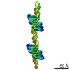


 PDBj
PDBj


 Assembly
 Sample preparation
 Electron microscopy imaging
 FIELD EMISSION GUN / Accelerating voltage: 300 kV / Illumination mode: FLOOD BEAM
 Processing