[English] 日本語
 Yorodumi
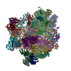
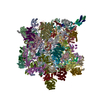
- PDB-6yxx: State A of the Trypanosoma brucei mitoribosomal large subunit ass... -
+ Open data
- Basic information
| Entry | Database: PDB / ID: 6yxx | ||||||
|---|---|---|---|---|---|---|---|
| Title | State A of the Trypanosoma brucei mitoribosomal large subunit assembly intermediate | ||||||
 Components Components |
| ||||||
 Keywords Keywords | RIBOSOME / mitoribosome / assembly / LSU | ||||||
| Function / homology |  Function and homology information Function and homology informationorganellar large ribosomal subunit / pseudouridine synthesis / pseudouridine synthase activity / kinetoplast / RNA methyltransferase activity / nuclear lumen / mitochondrial large ribosomal subunit / ciliary plasm / mitochondrial ribosome / mitochondrial translation ...organellar large ribosomal subunit / pseudouridine synthesis / pseudouridine synthase activity / kinetoplast / RNA methyltransferase activity / nuclear lumen / mitochondrial large ribosomal subunit / ciliary plasm / mitochondrial ribosome / mitochondrial translation / post-transcriptional regulation of gene expression / cyclosporin A binding / : / acyl binding / acyl carrier activity / RNA processing / peptidylprolyl isomerase / peptidyl-prolyl cis-trans isomerase activity / lipid metabolic process / fatty acid biosynthetic process / large ribosomal subunit / protein folding / large ribosomal subunit rRNA binding / RNA helicase activity / negative regulation of translation / rRNA binding / hydrolase activity / ribosome / structural constituent of ribosome / mitochondrial matrix / ribonucleoprotein complex / translation / GTPase activity / mRNA binding / GTP binding / nucleolus / mitochondrion / RNA binding / nucleoplasm / ATP binding / nucleus / cytosol / cytoplasm Similarity search - Function | ||||||
| Biological species |  | ||||||
| Method | ELECTRON MICROSCOPY / single particle reconstruction / cryo EM / Resolution: 3.9 Å | ||||||
 Authors Authors | Jaskolowski, M. / Ramrath, D.J.F. / Bieri, P. / Niemann, M. / Mattei, S. / Calderaro, S. / Leibundgut, M.A. / Horn, E.K. / Boehringer, D. / Schneider, A. / Ban, N. | ||||||
| Funding support |  Switzerland, 1items Switzerland, 1items
| ||||||
 Citation Citation |  Journal: Mol Cell / Year: 2020 Journal: Mol Cell / Year: 2020Title: Structural Insights into the Mechanism of Mitoribosomal Large Subunit Biogenesis. Authors: Mateusz Jaskolowski / David J F Ramrath / Philipp Bieri / Moritz Niemann / Simone Mattei / Salvatore Calderaro / Marc Leibundgut / Elke K Horn / Daniel Boehringer / André Schneider / Nenad Ban /  Abstract: In contrast to the bacterial translation machinery, mitoribosomes and mitochondrial translation factors are highly divergent in terms of composition and architecture. There is increasing evidence ...In contrast to the bacterial translation machinery, mitoribosomes and mitochondrial translation factors are highly divergent in terms of composition and architecture. There is increasing evidence that the biogenesis of mitoribosomes is an intricate pathway, involving many assembly factors. To better understand this process, we investigated native assembly intermediates of the mitoribosomal large subunit from the human parasite Trypanosoma brucei using cryo-electron microscopy. We identify 28 assembly factors, 6 of which are homologous to bacterial and eukaryotic ribosome assembly factors. They interact with the partially folded rRNA by specifically recognizing functionally important regions such as the peptidyltransferase center. The architectural and compositional comparison of the assembly intermediates indicates a stepwise modular assembly process, during which the rRNA folds toward its mature state. During the process, several conserved GTPases and a helicase form highly intertwined interaction networks that stabilize distinct assembly intermediates. The presented structures provide general insights into mitoribosomal maturation. | ||||||
| History |
|
- Structure visualization
| Movie |
 Movie viewer Movie viewer |
|---|---|
| Structure viewer | Molecule:  Molmil Molmil Jmol/JSmol Jmol/JSmol |
- Downloads & links
- Download
| PDBx/mmCIF format |  6yxx.cif.gz 6yxx.cif.gz | 4.1 MB | Display |  PDBx/mmCIF format PDBx/mmCIF format |
|---|---|---|---|---|
| PDB format |  pdb6yxx.ent.gz pdb6yxx.ent.gz | Display |  PDB format PDB format | |
| PDBx/mmJSON format |  6yxx.json.gz 6yxx.json.gz | Tree view |  PDBx/mmJSON format PDBx/mmJSON format | |
| Others |  Other downloads Other downloads |
-Validation report
| Summary document |  6yxx_validation.pdf.gz 6yxx_validation.pdf.gz | 2.3 MB | Display |  wwPDB validaton report wwPDB validaton report |
|---|---|---|---|---|
| Full document |  6yxx_full_validation.pdf.gz 6yxx_full_validation.pdf.gz | 2.3 MB | Display | |
| Data in XML |  6yxx_validation.xml.gz 6yxx_validation.xml.gz | 454.1 KB | Display | |
| Data in CIF |  6yxx_validation.cif.gz 6yxx_validation.cif.gz | 750.6 KB | Display | |
| Arichive directory |  https://data.pdbj.org/pub/pdb/validation_reports/yx/6yxx https://data.pdbj.org/pub/pdb/validation_reports/yx/6yxx ftp://data.pdbj.org/pub/pdb/validation_reports/yx/6yxx ftp://data.pdbj.org/pub/pdb/validation_reports/yx/6yxx | HTTPS FTP |
-Related structure data
| Related structure data |  10999MC  6yxyC M: map data used to model this data C: citing same article ( |
|---|---|
| Similar structure data |
- Links
Links
- Assembly
| Deposited unit | 
|
|---|---|
| 1 |
|
- Components
+Protein , 68 types, 69 molecules A1E1A2E2A3E3E4A5E5E6A8BAEABBEBECBDEDBEBFEGBHEHAIBIBJBKBLELEM...
-RNA chain , 1 types, 1 molecules AA
| #12: RNA chain | Mass: 365178.281 Da / Num. of mol.: 1 / Source method: isolated from a natural source / Source: (natural)  |
|---|
-Protein/peptide , 6 types, 8 molecules UAUfUIUrUKUMUmUn
| #15: Protein/peptide | Mass: 869.063 Da / Num. of mol.: 2 / Source method: isolated from a natural source / Source: (natural)  #32: Protein/peptide | Mass: 1805.216 Da / Num. of mol.: 2 / Source method: isolated from a natural source / Source: (natural)  #36: Protein/peptide | | Mass: 2145.636 Da / Num. of mol.: 1 / Source method: isolated from a natural source / Source: (natural)  #40: Protein/peptide | | Mass: 698.854 Da / Num. of mol.: 1 / Source method: isolated from a natural source / Source: (natural)  #77: Protein/peptide | | Mass: 2315.846 Da / Num. of mol.: 1 / Source method: isolated from a natural source / Source: (natural)  #78: Protein/peptide | | Mass: 2400.951 Da / Num. of mol.: 1 / Source method: isolated from a natural source / Source: (natural)  |
|---|
-Ribosomal protein ... , 3 types, 3 molecules AEAFAK
| #21: Protein | Mass: 55073.699 Da / Num. of mol.: 1 / Source method: isolated from a natural source / Source: (natural)  |
|---|---|
| #24: Protein | Mass: 53156.664 Da / Num. of mol.: 1 / Source method: isolated from a natural source / Source: (natural)  |
| #34: Protein | Mass: 40266.777 Da / Num. of mol.: 1 / Source method: isolated from a natural source / Source: (natural)  |
-SpoU methylase domain-containing ... , 2 types, 2 molecules EEEF
| #23: Protein | Mass: 67167.289 Da / Num. of mol.: 1 / Source method: isolated from a natural source / Source: (natural)  |
|---|---|
| #26: Protein | Mass: 40223.109 Da / Num. of mol.: 1 / Source method: isolated from a natural source / Source: (natural)  |
-50S ribosomal protein ... , 2 types, 2 molecules ANAR
| #41: Protein | Mass: 24341.309 Da / Num. of mol.: 1 / Source method: isolated from a natural source / Source: (natural)  |
|---|---|
| #48: Protein | Mass: 35117.824 Da / Num. of mol.: 1 / Source method: isolated from a natural source / Source: (natural)  |
-Peptidyl-prolyl cis-trans ... , 2 types, 2 molecules BQBZ
| #47: Protein | Mass: 25579.004 Da / Num. of mol.: 1 / Source method: isolated from a natural source / Source: (natural)  |
|---|---|
| #64: Protein | Mass: 20542.291 Da / Num. of mol.: 1 / Source method: isolated from a natural source / Source: (natural)  |
-Non-polymers , 8 types, 39 molecules 


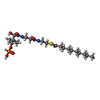
| #85: Chemical | ChemComp-ZN / #86: Chemical | ChemComp-MG / #87: Chemical | #88: Chemical | #89: Chemical | ChemComp-ATP / | #90: Chemical | ChemComp-PM8 / | #91: Chemical | ChemComp-NAD / | #92: Water | ChemComp-HOH / | |
|---|
-Details
| Has ligand of interest | N |
|---|---|
| Has protein modification | Y |
-Experimental details
-Experiment
| Experiment | Method: ELECTRON MICROSCOPY |
|---|---|
| EM experiment | Aggregation state: PARTICLE / 3D reconstruction method: single particle reconstruction |
- Sample preparation
| Component | Name: State B of the Trypanosoma brucei mitoribosomal large subunit assembly intermediate Type: RIBOSOME / Entity ID: #1-#84 / Source: NATURAL |
|---|---|
| Source (natural) | Organism:  |
| Buffer solution | pH: 7.4 |
| Specimen | Embedding applied: NO / Shadowing applied: NO / Staining applied: NO / Vitrification applied: YES |
| Specimen support | Grid type: Quantifoil R2/2 |
| Vitrification | Instrument: FEI VITROBOT MARK IV / Cryogen name: ETHANE-PROPANE / Humidity: 100 % / Chamber temperature: 277 K Details: 3.5 uL of the sample was blotted for 7-9 sec before plunging |
- Electron microscopy imaging
| Experimental equipment |  Model: Titan Krios / Image courtesy: FEI Company |
|---|---|
| Microscopy | Model: FEI TITAN KRIOS |
| Electron gun | Electron source:  FIELD EMISSION GUN / Accelerating voltage: 300 kV / Illumination mode: FLOOD BEAM FIELD EMISSION GUN / Accelerating voltage: 300 kV / Illumination mode: FLOOD BEAM |
| Electron lens | Mode: BRIGHT FIELD / Cs: 2.7 mm / C2 aperture diameter: 100 µm / Alignment procedure: COMA FREE |
| Specimen holder | Cryogen: NITROGEN / Specimen holder model: FEI TITAN KRIOS AUTOGRID HOLDER |
| Image recording | Electron dose: 75 e/Å2 / Detector mode: INTEGRATING / Film or detector model: FEI FALCON III (4k x 4k) |
- Processing
| EM software | Name: RELION / Version: 3 / Category: 3D reconstruction |
|---|---|
| CTF correction | Type: PHASE FLIPPING AND AMPLITUDE CORRECTION |
| Symmetry | Point symmetry: C1 (asymmetric) |
| 3D reconstruction | Resolution: 3.9 Å / Resolution method: FSC 0.143 CUT-OFF / Num. of particles: 16215 / Symmetry type: POINT |
| Atomic model building | Details: Initial models of ribosomal proteins of the T. brucei mitochondrial ribosomal large subunit were taken from PDB 6HIX. Assembly factors of the mitoribosomal large subunit were built de novo. |
 Movie
Movie Controller


 PDBj
PDBj


