Movie
Movie Controller

[English] 日本語
 Yorodumi
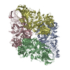
- PDB-6x1q: 1.8 Angstrom resolution structure of b-galactosidase with a 200 k... -
+ Open data

- Basic information
| Entry | Database: PDB / ID: 6x1q | ||||||
|---|---|---|---|---|---|---|---|
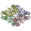
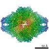
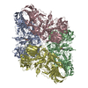
| Title | 1.8 Angstrom resolution structure of b-galactosidase with a 200 kV cryoARM electron microscope | ||||||
 Components Components | Beta-galactosidase | ||||||
 Keywords Keywords | HYDROLASE / enzyme | ||||||
| Function / homology |  Function and homology information Function and homology informationalkali metal ion binding / lactose catabolic process / beta-galactosidase complex / beta-galactosidase / beta-galactosidase activity / carbohydrate binding / magnesium ion binding / identical protein binding Similarity search - Function | ||||||
| Biological species |  | ||||||
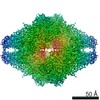
| Method | ELECTRON MICROSCOPY / single particle reconstruction / cryo EM / Resolution: 1.8 Å | ||||||
 Authors Authors | Merk, A. / Fukumura, T. / Zhu, X. / Darling, J. / Grisshammer, R. / Ognjenovic, J. / Subramaniam, S. | ||||||
| Funding support | 1items
| ||||||
 Citation Citation | Journal: IUCrJ / Year: 2020 Title: 1.8 Å resolution structure of β-galactosidase with a 200 kV CRYO ARM electron microscope. Authors: Alan Merk / Takuma Fukumura / Xing Zhu / Joseph E Darling / Reinhard Grisshammer / Jana Ognjenovic / Sriram Subramaniam /    Abstract: We report the determination of the structure of β-galactosidase at a resolution of ∼1.8 Å using data collected on a 200 kV CRYO ARM microscope equipped with a K3 direct electron detector. ...We report the determination of the structure of β-galactosidase at a resolution of ∼1.8 Å using data collected on a 200 kV CRYO ARM microscope equipped with a K3 direct electron detector. The data were collected in a single 24 h session by recording images from an array of 7 × 7 holes at each stage position using the automated data collection program . In addition to the expected features such as holes in the densities of aromatic residues, the map also shows density bumps corresponding to the locations of hydrogen atoms. The hydrogen densities are useful in assigning absolute orientations for residues such as glutamine or asparagine by removing the uncertainty in the fitting of the amide groups, and are likely to be especially relevant in the context of structure-guided drug design. These findings validate the use of electron microscopes operating at 200 kV for imaging protein complexes at atomic resolution using cryo-EM. | ||||||
| History |
|
- Structure visualization
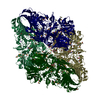
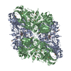
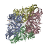
| Movie |
Movie viewer |
|---|---|
| Structure viewer | Molecule: MolmilJmol/JSmol |
- Downloads & links
-Download
| PDBx/mmCIF format | 6x1q.cif.gz | 1.4 MB | Display | PDBx/mmCIF format |
|---|---|---|---|---|
| PDB format | pdb6x1q.ent.gz | 1.1 MB | Display | PDB format |
| PDBx/mmJSON format | 6x1q.json.gz | Tree view | PDBx/mmJSON format | |
| Others |  Other downloads Other downloads |
-Validation report
| Arichive directory | https://data.pdbj.org/pub/pdb/validation_reports/x1/6x1qftp://data.pdbj.org/pub/pdb/validation_reports/x1/6x1q | HTTPS FTP |
|---|
-Related structure data
| Related structure data |  21995MC M: map data used to model this data C: citing same article ( |
|---|---|
| Similar structure data | |
| EM raw data | EMPIAR-10446 (Title: 1.8 Å resolution structure of β-galactosidase with a 200 kV CRYO ARM electron microscope Data size: 1.1 TB Data #1: uncorrected multi-frame micrographs of beta-galactosidase [micrographs - multiframe]) |

-Links
 PDBj
PDBj


- Assembly
| Deposited unit | 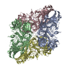
|
|---|---|
| 1 |
|
-Components
| #1: Protein | Mass: 116181.031 Da / Num. of mol.: 4 / Source method: isolated from a natural source / Source: (natural) #2: Chemical | ChemComp-MG /   Mass: 24.305 Da / Num. of mol.: 8 / Source method: obtained synthetically / Formula: Mg Mass: 24.305 Da / Num. of mol.: 8 / Source method: obtained synthetically / Formula: Mg#3: Chemical | ChemComp-NA /   Mass: 22.990 Da / Num. of mol.: 8 / Source method: obtained synthetically / Formula: Na Mass: 22.990 Da / Num. of mol.: 8 / Source method: obtained synthetically / Formula: Na#4: Water | ChemComp-HOH / |  Mass: 18.015 Da / Num. of mol.: 3618 / Source method: isolated from a natural source / Formula: H2O Mass: 18.015 Da / Num. of mol.: 3618 / Source method: isolated from a natural source / Formula: H2OHas ligand of interest | N | |
|---|
-Experimental details
-Experiment
| Experiment | Method: ELECTRON MICROSCOPY |
|---|---|
| EM experiment | Aggregation state: PARTICLE / 3D reconstruction method: single particle reconstruction |
- Sample preparation
| Component | Name: beta-galactosidase / Type: COMPLEX / Entity ID: #1 / Source: RECOMBINANT |
|---|---|
| Molecular weight | Value: .465 MDa / Experimental value: NO |
| Source (natural) | Organism: |
| Source (recombinant) | Organism: |
| Buffer solution | pH: 8 / Details: 25mM Tris pH 8.0, 50mM NaCl, 0.5mM TCEP, 2mM MgCl |
| Specimen | Conc.: 4.5 mg/ml / Embedding applied: NO / Shadowing applied: NO / Staining applied: NO / Vitrification applied: YES / Details: Purified by size exclusion chromatography |
| Specimen support | Grid material: COPPER / Grid mesh size: 200 divisions/in. / Grid type: Quantifoil R1.2/1.3 |
| Vitrification | Instrument: LEICA EM GP / Cryogen name: ETHANE / Humidity: 95 % / Chamber temperature: 291 K |
- Electron microscopy imaging
| Microscopy | Model: JEOL CRYO ARM 200 |
|---|---|
| Electron gun | Electron source:  FIELD EMISSION GUN / Accelerating voltage: 200 kV / Illumination mode: FLOOD BEAM FIELD EMISSION GUN / Accelerating voltage: 200 kV / Illumination mode: FLOOD BEAM |
| Electron lens | Mode: BRIGHT FIELD / Nominal defocus max: -950 nm / Nominal defocus min: -800 nm / Cs: 2.7 mm / C2 aperture diameter: 70 µm / Alignment procedure: COMA FREE |
| Specimen holder | Cryogen: NITROGEN / Specimen holder model: JEOL CRYOSPECPORTER |
| Image recording | Average exposure time: 1 sec. / Electron dose: 40 e/Å2 / Film or detector model: GATAN K3 (6k x 4k) / Num. of grids imaged: 1 / Num. of real images: 4949 |
| EM imaging optics | Energyfilter name: In-column Omega Filter / Energyfilter slit width: 30 eV |
- Processing
| Software | Name: PHENIX / Version: 1.17.1_3660: / Classification: refinement | ||||||||||||||||||||||||||||
|---|---|---|---|---|---|---|---|---|---|---|---|---|---|---|---|---|---|---|---|---|---|---|---|---|---|---|---|---|---|
| EM software |
| ||||||||||||||||||||||||||||
| CTF correction | Type: PHASE FLIPPING AND AMPLITUDE CORRECTION | ||||||||||||||||||||||||||||
| Particle selection | Num. of particles selected: 998945 | ||||||||||||||||||||||||||||
| Symmetry | Point symmetry: D2 (2x2 fold dihedral) | ||||||||||||||||||||||||||||
| 3D reconstruction | Resolution: 1.8 Å / Resolution method: FSC 0.143 CUT-OFF / Num. of particles: 257202 / Symmetry type: POINT | ||||||||||||||||||||||||||||
| Atomic model building | PDB-ID: 5A1A Accession code: 5A1A / Source name: PDB / Type: experimental model | ||||||||||||||||||||||||||||
| Refine LS restraints |
|