 Movie
Movie Controller


+ Open data

- Basic information
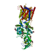
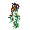
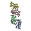
| Entry | Database: PDB / ID: 6w5s | ||||||||||||||||||||||||||||||||||||||||||
|---|---|---|---|---|---|---|---|---|---|---|---|---|---|---|---|---|---|---|---|---|---|---|---|---|---|---|---|---|---|---|---|---|---|---|---|---|---|---|---|---|---|---|---|
| Title | NPC1 structure in GDN micelles at pH 8.0 | ||||||||||||||||||||||||||||||||||||||||||
 Components Components | NPC intracellular cholesterol transporter 1 | ||||||||||||||||||||||||||||||||||||||||||
 Keywords Keywords | TRANSPORT PROTEIN / Cholesterol Lysosome | ||||||||||||||||||||||||||||||||||||||||||
| Function / homology |  Function and homology information Function and homology informationmembrane raft organization / cytoplasmic side of lysosomal membrane / intracellular cholesterol transport / lysosome to ER cholesterol transport / intracellular lipid transport / sterol transport / negative regulation of epithelial cell apoptotic process / intestinal cholesterol absorption / cholesterol transport / LDL clearance ...membrane raft organization / cytoplasmic side of lysosomal membrane / intracellular cholesterol transport / lysosome to ER cholesterol transport / intracellular lipid transport / sterol transport / negative regulation of epithelial cell apoptotic process / intestinal cholesterol absorption / cholesterol transport / LDL clearance / bile acid metabolic process / cholesterol transfer activity / glycoprotein biosynthetic process / establishment of protein localization to membrane / cholesterol efflux / lysosomal transport / cholesterol binding / response to cadmium ion / cholesterol metabolic process / negative regulation of TORC1 signaling / cholesterol homeostasis / autophagy / gene expression / transmembrane signaling receptor activity / nuclear envelope / late endosome membrane / virus receptor activity / signaling receptor activity / lysosome / membrane raft / lysosomal membrane / symbiont entry into host cell / perinuclear region of cytoplasm / Golgi apparatus / endoplasmic reticulum / extracellular exosome / extracellular region / membrane / plasma membrane Similarity search - Function | ||||||||||||||||||||||||||||||||||||||||||
| Biological species |  Homo sapiens (human) Homo sapiens (human) | ||||||||||||||||||||||||||||||||||||||||||
| Method | ELECTRON MICROSCOPY / single particle reconstruction / cryo EM / Resolution: 3 Å | ||||||||||||||||||||||||||||||||||||||||||
 Authors Authors | Yan, N. / Qian, H.W. / Wu, X.L. | ||||||||||||||||||||||||||||||||||||||||||
| Funding support |  United States, 1items United States, 1items
| ||||||||||||||||||||||||||||||||||||||||||
 Citation Citation | Journal: Cell / Year: 2020 Title: Structural Basis of Low-pH-Dependent Lysosomal Cholesterol Egress by NPC1 and NPC2. Authors: Hongwu Qian / Xuelan Wu / Ximing Du / Xia Yao / Xin Zhao / Joyce Lee / Hongyuan Yang / Nieng Yan /   Abstract: Lysosomal cholesterol egress requires two proteins, NPC1 and NPC2, whose defects are responsible for Niemann-Pick disease type C (NPC). Here, we present systematic structural characterizations that ...Lysosomal cholesterol egress requires two proteins, NPC1 and NPC2, whose defects are responsible for Niemann-Pick disease type C (NPC). Here, we present systematic structural characterizations that reveal the molecular basis for low-pH-dependent cholesterol delivery from NPC2 to the transmembrane (TM) domain of NPC1. At pH 8.0, similar structures of NPC1 were obtained in nanodiscs and in detergent at resolutions of 3.6 Å and 3.0 Å, respectively. A tunnel connecting the N-terminal domain (NTD) and the transmembrane sterol-sensing domain (SSD) was unveiled. At pH 5.5, the NTD exhibits two conformations, suggesting the motion for cholesterol delivery to the tunnel. A putative cholesterol molecule is found at the membrane boundary of the tunnel, and TM2 moves toward formation of a surface pocket on the SSD. Finally, the structure of the NPC1-NPC2 complex at 4.0 Å resolution was obtained at pH 5.5, elucidating the molecular basis for cholesterol handoff from NPC2 to NPC1(NTD). | ||||||||||||||||||||||||||||||||||||||||||
| History |
|
- Structure visualization
| Movie |
Movie viewer |
|---|---|
| Structure viewer | Molecule: MolmilJmol/JSmol |
- Downloads & links
-Download
| PDBx/mmCIF format | 6w5s.cif.gz | 286.2 KB | Display | PDBx/mmCIF format |
|---|---|---|---|---|
| PDB format | pdb6w5s.ent.gz | 216.3 KB | Display | PDB format |
| PDBx/mmJSON format | 6w5s.json.gz | Tree view | PDBx/mmJSON format | |
| Others |  Other downloads Other downloads |
-Validation report
| Arichive directory | https://data.pdbj.org/pub/pdb/validation_reports/w5/6w5sftp://data.pdbj.org/pub/pdb/validation_reports/w5/6w5s | HTTPS FTP |
|---|
-Related structure data
| Related structure data |  21546MC  6w5rC  6w5tC  6w5uC  6w5vC M: map data used to model this data C: citing same article ( |
|---|---|
| Similar structure data |


-Links
 PDBj
PDBj


- Assembly
| Deposited unit | 
|
|---|---|
| 1 |
|
-Components
-Protein , 1 types, 1 molecules A
| #1: Protein | Mass: 146083.688 Da / Num. of mol.: 1 Source method: isolated from a genetically manipulated source Source: (gene. exp.) Homo sapiens (human) / Gene: NPC1 / Production host: Homo sapiens (human) / References: UniProt: O15118 |
|---|
-Sugars , 4 types, 13 molecules 
| #2: Polysaccharide | Source method: isolated from a genetically manipulated source #3: Polysaccharide | alpha-D-mannopyranose-(1-3)-2-acetamido-2-deoxy-beta-D-glucopyranose-(1-4)-2-acetamido-2-deoxy-beta- ...alpha-D-mannopyranose-(1-3)-2-acetamido-2-deoxy-beta-D-glucopyranose-(1-4)-2-acetamido-2-deoxy-beta-D-glucopyranose | Source method: isolated from a genetically manipulated source #4: Polysaccharide | alpha-D-mannopyranose-(1-4)-2-acetamido-2-deoxy-beta-D-glucopyranose-(1-4)-2-acetamido-2-deoxy-beta- ...alpha-D-mannopyranose-(1-4)-2-acetamido-2-deoxy-beta-D-glucopyranose-(1-4)-2-acetamido-2-deoxy-beta-D-glucopyranose | Source method: isolated from a genetically manipulated source #5: Sugar | ChemComp-NAG /  Type: D-saccharide, beta linking / Mass: 221.208 Da / Num. of mol.: 9 Type: D-saccharide, beta linking / Mass: 221.208 Da / Num. of mol.: 9Source method: isolated from a genetically manipulated source Formula: C8H15NO6 |
|---|
-Non-polymers , 2 types, 2 molecules 

| #6: Chemical | ChemComp-CLR /  Mass: 386.654 Da / Num. of mol.: 1 / Source method: obtained synthetically / Formula: C27H46O / Feature type: SUBJECT OF INVESTIGATION Mass: 386.654 Da / Num. of mol.: 1 / Source method: obtained synthetically / Formula: C27H46O / Feature type: SUBJECT OF INVESTIGATION |
|---|---|
| #7: Chemical | ChemComp-POV / ( Mass: 760.076 Da / Num. of mol.: 1 / Source method: obtained synthetically / Formula: C42H82NO8P / Comment: phospholipid*YM Mass: 760.076 Da / Num. of mol.: 1 / Source method: obtained synthetically / Formula: C42H82NO8P / Comment: phospholipid*YM |
-Details
| Has ligand of interest | Y |
|---|---|
| Has protein modification | Y |
-Experimental details
-Experiment
| Experiment | Method: ELECTRON MICROSCOPY |
|---|---|
| EM experiment | Aggregation state: PARTICLE / 3D reconstruction method: single particle reconstruction |
- Sample preparation
| Component | Name: NPC1 at pH 8.0 / Type: COMPLEX / Entity ID: #1 / Source: RECOMBINANT |
|---|---|
| Molecular weight | Value: 0.15 MDa / Experimental value: NO |
| Source (natural) | Organism: Homo sapiens (human) |
| Source (recombinant) | Organism: Homo sapiens (human) |
| Buffer solution | pH: 8 |
| Specimen | Conc.: 15 mg/ml / Embedding applied: NO / Shadowing applied: NO / Staining applied: NO / Vitrification applied: YES |
| Specimen support | Details: unspecified |
| Vitrification | Cryogen name: ETHANE |
- Electron microscopy imaging
| Experimental equipment |  Model: Titan Krios / Image courtesy: FEI Company |
|---|---|
| Microscopy | Model: FEI TITAN KRIOS |
| Electron gun | Electron source:  FIELD EMISSION GUN / Accelerating voltage: 300 kV / Illumination mode: FLOOD BEAM FIELD EMISSION GUN / Accelerating voltage: 300 kV / Illumination mode: FLOOD BEAM |
| Electron lens | Mode: BRIGHT FIELD |
| Image recording | Electron dose: 50 e/Å2 / Film or detector model: GATAN K2 SUMMIT (4k x 4k) |
- Processing
| Software | Name: PHENIX / Version: 1.16_3549: / Classification: refinement | ||||||||||||||||||||||||
|---|---|---|---|---|---|---|---|---|---|---|---|---|---|---|---|---|---|---|---|---|---|---|---|---|---|
| EM software |
| ||||||||||||||||||||||||
| CTF correction | Type: PHASE FLIPPING AND AMPLITUDE CORRECTION | ||||||||||||||||||||||||
| 3D reconstruction | Resolution: 3 Å / Resolution method: FSC 0.143 CUT-OFF / Num. of particles: 813922 / Symmetry type: POINT | ||||||||||||||||||||||||
| Refine LS restraints |
|
