 Movie
Movie Controller

[English] 日本語
 Yorodumi
- PDB-6urt: Extended Sensor Paddles with Bound Lipids Revealed in Mechanosens... -
+ Open data

- Basic information
| Entry | Database: PDB / ID: 6urt | ||||||
|---|---|---|---|---|---|---|---|
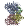
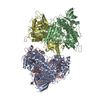
| Title | Extended Sensor Paddles with Bound Lipids Revealed in Mechanosensitive Channel YnaI | ||||||
 Components Components | Low conductance mechanosensitive channel YnaI | ||||||
 Keywords Keywords | MEMBRANE PROTEIN / mechanosensitive channel / Phosphoserine lipid / complete paddle | ||||||
| Function / homology |  Function and homology information Function and homology informationmechanosensitive monoatomic ion channel activity / cellular response to osmotic stress / monoatomic ion channel activity / identical protein binding / plasma membrane Similarity search - Function | ||||||
| Biological species |  | ||||||
| Method | ELECTRON MICROSCOPY / single particle reconstruction / cryo EM / Resolution: 3.27 Å | ||||||
 Authors Authors | Hu, W. / Wang, Z. / Zheng, H. | ||||||
 Citation Citation | Journal: Commun Biol / Year: 2021 Title: Mechanosensitive channel YnaI has lipid-bound extended sensor paddles. Authors: Wenxin Hu / Zhiming Wang / Hongjin Zheng /  Abstract: The general mechanism of bacterial mechanosensitive channels (MS) has been characterized by extensive studies on a small conductance channel MscS from Escherichia coli (E. coli). However, recent ...The general mechanism of bacterial mechanosensitive channels (MS) has been characterized by extensive studies on a small conductance channel MscS from Escherichia coli (E. coli). However, recent structural studies on the same channel have revealed controversial roles of various channel-bound lipids in channel gating. To better understand bacterial MscS-like channels, it is necessary to characterize homologs other than MscS. Here, we describe the structure of YnaI, one of the closest MscS homologs in E. coli, in its non-conducting state at 3.3 Å resolution determined by cryo electron microscopy. Our structure revealed the intact membrane sensor paddle domain in YnaI, which was stabilized by functionally important residues H43, Q46, Y50 and K93. In the pockets between sensor paddles, there were clear lipid densities that interact strongly with residues Q100 and R120. These lipids were a mixture of natural lipids but may be enriched in cardiolipin and phosphatidylserine. In addition, residues along the ion-conducting pathway and responsible for the heptameric assembly were discussed. Together with biochemical experiments and mutagenesis studies, our results provide strong support for the idea that the pocket lipids are functionally important for mechanosensitive channels. | ||||||
| History |
|
- Structure visualization
| Movie |
Movie viewer |
|---|---|
| Structure viewer | Molecule: MolmilJmol/JSmol |
- Downloads & links
-Download
| PDBx/mmCIF format | 6urt.cif.gz | 394.3 KB | Display | PDBx/mmCIF format |
|---|---|---|---|---|
| PDB format | pdb6urt.ent.gz | 329 KB | Display | PDB format |
| PDBx/mmJSON format | 6urt.json.gz | Tree view | PDBx/mmJSON format | |
| Others |  Other downloads Other downloads |
-Validation report
| Arichive directory | https://data.pdbj.org/pub/pdb/validation_reports/ur/6urtftp://data.pdbj.org/pub/pdb/validation_reports/ur/6urt | HTTPS FTP |
|---|
-Related structure data
| Related structure data | 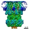 20862MC M: map data used to model this data C: citing same article ( |
|---|---|
| Similar structure data |


-Links
 PDBj
PDBj


- Assembly
| Deposited unit | 
|
|---|---|
| 1 |
|
-Components
| #1: Protein | Mass: 38792.344 Da / Num. of mol.: 7 Source method: isolated from a genetically manipulated source Source: (gene. exp.) Strain: K12 / Gene: ynaI, b1330, JW1323 / Production host: #2: Chemical | ChemComp-QGD /   Mass: 750.038 Da / Num. of mol.: 7 / Source method: obtained synthetically / Formula: C40H80NO9P Mass: 750.038 Da / Num. of mol.: 7 / Source method: obtained synthetically / Formula: C40H80NO9PHas ligand of interest | N | |
|---|
-Experimental details
-Experiment
| Experiment | Method: ELECTRON MICROSCOPY |
|---|---|
| EM experiment | Aggregation state: PARTICLE / 3D reconstruction method: single particle reconstruction |
- Sample preparation
| Component | Name: YnaI with PS lipids / Type: COMPLEX / Entity ID: #1 / Source: RECOMBINANT |
|---|---|
| Molecular weight | Value: 0.27 MDa / Experimental value: NO |
| Source (natural) | Organism: |
| Source (recombinant) | Organism: |
| Buffer solution | pH: 7.5 Details: 20mM HEPES pH7.5, 150mM NaCl, 0.01% LMNG, 3mM Fluorinated Fos-Cholin 8 |
| Specimen | Conc.: 5 mg/ml / Embedding applied: NO / Shadowing applied: NO / Staining applied: NO / Vitrification applied: YES |
| Specimen support | Grid material: COPPER / Grid mesh size: 400 divisions/in. / Grid type: C-flat-1.2/1.3 4C |
| Vitrification | Instrument: FEI VITROBOT MARK IV / Cryogen name: ETHANE / Humidity: 100 % / Chamber temperature: 277 K |
- Electron microscopy imaging
| Experimental equipment |  Model: Talos Arctica / Image courtesy: FEI Company |
|---|---|
| Microscopy | Model: FEI TECNAI ARCTICA |
| Electron gun | Electron source:  FIELD EMISSION GUN / Accelerating voltage: 200 kV / Illumination mode: FLOOD BEAM FIELD EMISSION GUN / Accelerating voltage: 200 kV / Illumination mode: FLOOD BEAM |
| Electron lens | Mode: BRIGHT FIELD / Cs: 2.7 mm / C2 aperture diameter: 70 µm |
| Specimen holder | Cryogen: NITROGEN / Specimen holder model: FEI TITAN KRIOS AUTOGRID HOLDER |
| Image recording | Average exposure time: 2 sec. / Electron dose: 64.3 e/Å2 / Film or detector model: GATAN K3 (6k x 4k) |
- Processing
| CTF correction | Type: PHASE FLIPPING ONLY |
|---|---|
| 3D reconstruction | Resolution: 3.27 Å / Resolution method: FSC 0.143 CUT-OFF / Num. of particles: 178000 / Symmetry type: POINT |
