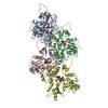
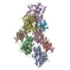
Movie
Movie Controller


+ Open data

- Basic information
| Entry | Database: PDB / ID: 6uc4 | |||||||||
|---|---|---|---|---|---|---|---|---|---|---|
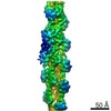
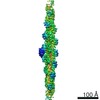
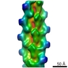
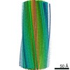
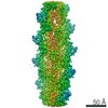
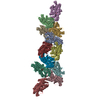
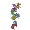
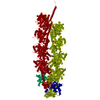
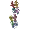
| Title | Barbed end side of a cofilactin cluster | |||||||||
 Components Components |
| |||||||||
 Keywords Keywords | STRUCTURAL PROTEIN / Cytoskeleton | |||||||||
| Function / homology |  Function and homology information Function and homology informationcellular response to ether / cofilin-actin rod / positive regulation of protein localization to cell leading edge / positive regulation of establishment of cell polarity regulating cell shape / negative regulation of unidimensional cell growth / positive regulation of barbed-end actin filament capping / negative regulation of lamellipodium assembly / negative regulation of postsynaptic density organization / actin filament fragmentation / positive regulation of actin filament depolymerization ...cellular response to ether / cofilin-actin rod / positive regulation of protein localization to cell leading edge / positive regulation of establishment of cell polarity regulating cell shape / negative regulation of unidimensional cell growth / positive regulation of barbed-end actin filament capping / negative regulation of lamellipodium assembly / negative regulation of postsynaptic density organization / actin filament fragmentation / positive regulation of actin filament depolymerization / negative regulation of actin filament bundle assembly / modification of postsynaptic actin cytoskeleton / positive regulation of embryonic development / negative regulation of actin filament depolymerization / positive regulation of cell motility / actin filament severing / host-mediated activation of viral process / negative regulation of cell adhesion / positive regulation of synaptic plasticity / establishment of spindle localization / regulation of dendritic spine morphogenesis / negative regulation of cell motility / actin filament depolymerization / RHO GTPases Activate ROCKs / cellular response to interleukin-6 / negative regulation of dendritic spine maintenance / cellular response to insulin-like growth factor stimulus / cytoskeletal motor activator activity / cortical actin cytoskeleton / positive regulation of dendritic spine development / phosphatidylinositol bisphosphate binding / myosin heavy chain binding / tropomyosin binding / actin filament bundle / troponin I binding / filamentous actin / mesenchyme migration / positive regulation of proteolysis / lamellipodium membrane / mitotic cytokinesis / skeletal muscle myofibril / striated muscle thin filament / cellular response to interleukin-1 / Sema3A PAK dependent Axon repulsion / actin filament bundle assembly / skeletal muscle thin filament assembly / Rho protein signal transduction / positive regulation of focal adhesion assembly / actin monomer binding / positive regulation of lamellipodium assembly / postsynaptic density, intracellular component / skeletal muscle fiber development / actin filament polymerization / stress fiber / titin binding / EPHB-mediated forward signaling / Gene and protein expression by JAK-STAT signaling after Interleukin-12 stimulation / cellular response to epidermal growth factor stimulus / protein import into nucleus / response to activity / hippocampus development / synaptic membrane / cellular response to tumor necrosis factor / filopodium / actin filament / Regulation of actin dynamics for phagocytic cup formation / response to virus / cellular response to hydrogen peroxide / Hydrolases; Acting on acid anhydrides; Acting on acid anhydrides to facilitate cellular and subcellular movement / ruffle membrane / mitochondrial membrane / nuclear matrix / calcium-dependent protein binding / cell-cell junction / actin filament binding / actin cytoskeleton organization / actin cytoskeleton / Platelet degranulation / positive regulation of cell growth / lamellipodium / growth cone / cell body / protein phosphatase binding / vesicle / dendritic spine / protein domain specific binding / signaling receptor binding / focal adhesion / hydrolase activity / neuronal cell body / positive regulation of gene expression / calcium ion binding / negative regulation of apoptotic process / glutamatergic synapse / magnesium ion binding / : / extracellular exosome / ATP binding / membrane / identical protein binding Similarity search - Function | |||||||||
| Biological species |  Homo sapiens (human) Homo sapiens (human)  Oryctolagus cuniculus (rabbit) Oryctolagus cuniculus (rabbit) | |||||||||
| Method | ELECTRON MICROSCOPY / single particle reconstruction / cryo EM / Resolution: 9.2 Å | |||||||||
 Authors Authors | Huehn, A.R. / Bibeau, J.P. / Schramm, A.C. / Cao, W. / De La Cruz, E.M. / Sindelar, C.V. | |||||||||
| Funding support |  United States, 2items United States, 2items
| |||||||||
 Citation Citation | Journal: Proc Natl Acad Sci U S A / Year: 2020 Title: Structures of cofilin-induced structural changes reveal local and asymmetric perturbations of actin filaments. Authors: Andrew R Huehn / Jeffrey P Bibeau / Anthony C Schramm / Wenxiang Cao / Enrique M De La Cruz / Charles V Sindelar / Abstract: Members of the cofilin/ADF family of proteins sever actin filaments, increasing the number of filament ends available for polymerization or depolymerization. Cofilin binds actin filaments with ...Members of the cofilin/ADF family of proteins sever actin filaments, increasing the number of filament ends available for polymerization or depolymerization. Cofilin binds actin filaments with positive cooperativity, forming clusters of contiguously bound cofilin along the filament lattice. Filament severing occurs preferentially at boundaries between bare and cofilin-decorated (cofilactin) segments and is biased at 1 side of a cluster. A molecular understanding of cooperative binding and filament severing has been impeded by a lack of structural data describing boundaries. Here, we apply methods for analyzing filament cryo-electron microscopy (cryo-EM) data at the single subunit level to directly investigate the structure of boundaries within partially decorated cofilactin filaments. Subnanometer resolution maps of isolated, bound cofilin molecules and an actin-cofilactin boundary indicate that cofilin-induced actin conformational changes are local and limited to subunits directly contacting bound cofilin. An isolated, bound cofilin compromises longitudinal filament contacts of 1 protofilament, consistent with a single cofilin having filament-severing activity. An individual, bound phosphomimetic (S3D) cofilin with weak severing activity adopts a unique binding mode that does not perturb actin structure. Cofilin clusters disrupt both protofilaments, consistent with a higher severing activity at boundaries compared to single cofilin. Comparison of these structures indicates that this disruption is substantially greater at pointed end sides of cofilactin clusters than at the barbed end. These structures, with the distribution of bound cofilin clusters, suggest that maximum binding cooperativity is achieved when 2 cofilins occupy adjacent sites. These results reveal the structural origins of cooperative cofilin binding and actin filament severing. | |||||||||
| History |
|
- Structure visualization
| Movie |
Movie viewer |
|---|---|
| Structure viewer | Molecule: MolmilJmol/JSmol |
- Downloads & links
-Download
| PDBx/mmCIF format | 6uc4.cif.gz | 696.7 KB | Display | PDBx/mmCIF format |
|---|---|---|---|---|
| PDB format | pdb6uc4.ent.gz | 578.4 KB | Display | PDB format |
| PDBx/mmJSON format | 6uc4.json.gz | Tree view | PDBx/mmJSON format | |
| Others |  Other downloads Other downloads |
-Validation report
| Arichive directory | https://data.pdbj.org/pub/pdb/validation_reports/uc/6uc4ftp://data.pdbj.org/pub/pdb/validation_reports/uc/6uc4 | HTTPS FTP |
|---|
-Related structure data
| Related structure data | 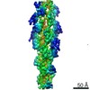 20726MC  6ubyC  6uc0C  6vaoC  6vauC C: citing same article ( M: map data used to model this data |
|---|---|
| Similar structure data |

-Links
 PDBj
PDBj







- Assembly
| Deposited unit | 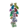
|
|---|---|
| 1 |
|
-Components
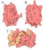
| #1: Protein | Mass: 42096.953 Da / Num. of mol.: 11 / Source method: isolated from a natural source / Source: (natural) Oryctolagus cuniculus (rabbit) / References: UniProt: P68135#2: Protein | Mass: 18532.531 Da / Num. of mol.: 5 Source method: isolated from a genetically manipulated source Source: (gene. exp.) Homo sapiens (human) / Gene: CFL1, CFL / Production host:   Escherichia coli (E. coli) / References: UniProt: P23528 Escherichia coli (E. coli) / References: UniProt: P23528#3: Chemical | ChemComp-MG /   Mass: 24.305 Da / Num. of mol.: 9 / Source method: obtained synthetically / Formula: Mg Mass: 24.305 Da / Num. of mol.: 9 / Source method: obtained synthetically / Formula: Mg#4: Chemical | ChemComp-ADP /  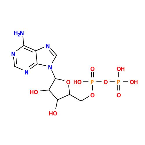 Mass: 427.201 Da / Num. of mol.: 9 / Source method: obtained synthetically / Formula: C10H15N5O10P2 / Comment: ADP, energy-carrying molecule*YM Mass: 427.201 Da / Num. of mol.: 9 / Source method: obtained synthetically / Formula: C10H15N5O10P2 / Comment: ADP, energy-carrying molecule*YMHas ligand of interest | N | Has protein modification | N | Sequence details | The molecules in the experiments were: Rabbit actin (UniProt P68135) and Human cofilin-1 (UniProt ...The molecules in the experiments were: Rabbit actin (UniProt P68135) and Human cofilin-1 (UniProt P23528), however the sequences modeled in the coordinates correspond to the sequences from Chicken actin and Chicken cofilin-2 since those were pdb models of homolog molecules used to fit into the cryo-EM density maps. | |
|---|
-Experimental details
-Experiment
| Experiment | Method: ELECTRON MICROSCOPY |
|---|---|
| EM experiment | Aggregation state: HELICAL ARRAY / 3D reconstruction method: single particle reconstruction |
- Sample preparation
| Component |
| ||||||||||||||||||||||||||||
|---|---|---|---|---|---|---|---|---|---|---|---|---|---|---|---|---|---|---|---|---|---|---|---|---|---|---|---|---|---|
| Molecular weight |
| ||||||||||||||||||||||||||||
| Source (natural) |
| ||||||||||||||||||||||||||||
| Source (recombinant) | Organism: Escherichia coli (E. coli) | ||||||||||||||||||||||||||||
| Buffer solution | pH: 6.6 | ||||||||||||||||||||||||||||
| Specimen | Embedding applied: NO / Shadowing applied: NO / Staining applied: NO / Vitrification applied: YES | ||||||||||||||||||||||||||||
| Vitrification | Instrument: HOMEMADE PLUNGER / Cryogen name: ETHANE |
- Electron microscopy imaging
| Experimental equipment |  Model: Titan Krios / Image courtesy: FEI Company |
|---|---|
| Microscopy | Model: FEI TITAN KRIOS |
| Electron gun | Electron source:  FIELD EMISSION GUN / Accelerating voltage: 300 kV / Illumination mode: SPOT SCAN FIELD EMISSION GUN / Accelerating voltage: 300 kV / Illumination mode: SPOT SCAN |
| Electron lens | Mode: BRIGHT FIELD |
| Image recording | Electron dose: 50 e/Å2 / Film or detector model: GATAN K2 SUMMIT (4k x 4k) |
- Processing
| EM software | Name: UCSF Chimera / Category: model fitting | ||||||||||||||||||||||||
|---|---|---|---|---|---|---|---|---|---|---|---|---|---|---|---|---|---|---|---|---|---|---|---|---|---|
| CTF correction | Type: PHASE FLIPPING AND AMPLITUDE CORRECTION | ||||||||||||||||||||||||
| Particle selection | Num. of particles selected: 1117338 Details: Both bare and cofilin-decorated segments were selected and initially refined together. | ||||||||||||||||||||||||
| 3D reconstruction | Resolution: 9.2 Å / Resolution method: FSC 0.143 CUT-OFF / Num. of particles: 671 / Symmetry type: POINT | ||||||||||||||||||||||||
| Atomic model building | 3D fitting-ID: 1 / Source name: PDB / Type: experimental model
|