 Movie
Movie Controller

[English] 日本語
 Yorodumi
- PDB-6ski: The Tle hydrolase bound to the TTR domain of the VgrG spike of th... -
+ Open data

- Basic information
| Entry | Database: PDB / ID: 6ski | |||||||||
|---|---|---|---|---|---|---|---|---|---|---|
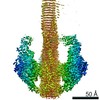
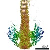
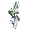
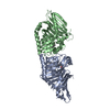
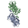
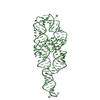
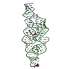
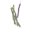
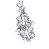
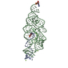
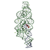
| Title | The Tle hydrolase bound to the TTR domain of the VgrG spike of the Type 6 secretion system | |||||||||
 Components Components | (Putative type VI secretion protein) x 2 | |||||||||
 Keywords Keywords | HYDROLASE / Lipase / effector / toxin | |||||||||
| Function / homology |  Function and homology information Function and homology informationDomain of unknown function DUF2235 / T6SS, Phospholipase effector Tle1-like, catalytic domain / Type VI secretion system spike protein VgrG2, C-terminal domain of unknown function DUF2345 / Putative type VI secretion system, Rhs element associated Vgr domain / Uncharacterized protein conserved in bacteria (DUF2345) / Putative type VI secretion system Rhs element Vgr / Type VI secretion system, RhsGE-associated Vgr family subset / Type VI secretion system, RhsGE-associated Vgr protein / Phage tail baseplate hub (GPD) / Gp5/Type VI secretion system Vgr protein, OB-fold domain ...Domain of unknown function DUF2235 / T6SS, Phospholipase effector Tle1-like, catalytic domain / Type VI secretion system spike protein VgrG2, C-terminal domain of unknown function DUF2345 / Putative type VI secretion system, Rhs element associated Vgr domain / Uncharacterized protein conserved in bacteria (DUF2345) / Putative type VI secretion system Rhs element Vgr / Type VI secretion system, RhsGE-associated Vgr family subset / Type VI secretion system, RhsGE-associated Vgr protein / Phage tail baseplate hub (GPD) / Gp5/Type VI secretion system Vgr protein, OB-fold domain / Type VI secretion system/phage-baseplate injector OB domain / Vgr protein, OB-fold domain superfamily Similarity search - Domain/homology | |||||||||
| Biological species |  | |||||||||
| Method | ELECTRON MICROSCOPY / single particle reconstruction / cryo EM / Resolution: 2.6 Å | |||||||||
 Authors Authors | Rapisarda, C. / Fronzes, R. | |||||||||
| Funding support |  France, France,  Germany, 2items Germany, 2items
| |||||||||
 Citation Citation | Journal: EMBO J / Year: 2020 Title: Structural basis for loading and inhibition of a bacterial T6SS phospholipase effector by the VgrG spike. Authors: Nicolas Flaugnatti / Chiara Rapisarda / Martial Rey / Solène G Beauvois / Viet Anh Nguyen / Stéphane Canaan / Eric Durand / Julia Chamot-Rooke / Eric Cascales / Rémi Fronzes / Laure Journet / Abstract: The bacterial type VI secretion system (T6SS) is a macromolecular machine that injects effectors into prokaryotic and eukaryotic cells. The mode of action of the T6SS is similar to contractile phages: ...The bacterial type VI secretion system (T6SS) is a macromolecular machine that injects effectors into prokaryotic and eukaryotic cells. The mode of action of the T6SS is similar to contractile phages: the contraction of a sheath structure pushes a tube topped by a spike into target cells. Effectors are loaded onto the spike or confined into the tube. In enteroaggregative Escherichia coli, the Tle1 phospholipase binds the C-terminal extension of the VgrG trimeric spike. Here, we purify the VgrG-Tle1 complex and show that a VgrG trimer binds three Tle1 monomers and inhibits their activity. Using covalent cross-linking coupled to high-resolution mass spectrometry, we provide information on the sites of contact and further identify the requirement for a Tle1 N-terminal secretion sequence in complex formation. Finally, we report the 2.6-Å-resolution cryo-electron microscopy tri-dimensional structure of the (VgrG) -(Tle1) complex revealing how the effector binds its cargo, and how VgrG inhibits Tle1 phospholipase activity. The inhibition of Tle1 phospholipase activity once bound to VgrG suggests that Tle1 dissociation from VgrG is required upon delivery. | |||||||||
| History |
|
- Structure visualization
| Movie |
Movie viewer |
|---|---|
| Structure viewer | Molecule: MolmilJmol/JSmol |
- Downloads & links
-Download
| PDBx/mmCIF format | 6ski.cif.gz | 127.3 KB | Display | PDBx/mmCIF format |
|---|---|---|---|---|
| PDB format | pdb6ski.ent.gz | 88.5 KB | Display | PDB format |
| PDBx/mmJSON format | 6ski.json.gz | Tree view | PDBx/mmJSON format | |
| Others |  Other downloads Other downloads |
-Validation report
| Summary document | 6ski_validation.pdf.gz | 1.1 MB | Display | wwPDB validaton report |
|---|---|---|---|---|
| Full document | 6ski_full_validation.pdf.gz | 1.1 MB | Display | |
| Data in XML | 6ski_validation.xml.gz | 30.5 KB | Display | |
| Data in CIF | 6ski_validation.cif.gz | 42 KB | Display | |
| Arichive directory | https://data.pdbj.org/pub/pdb/validation_reports/sk/6skiftp://data.pdbj.org/pub/pdb/validation_reports/sk/6ski | HTTPS FTP |
-Related structure data
| Related structure data | 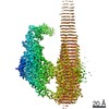 10225MC 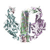 6sjlC 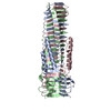 6sk0C M: map data used to model this data C: citing same article ( |
|---|---|
| Similar structure data |

-Links
 PDBj
PDBj- Assembly
| Deposited unit | 
|
|---|---|
| 1 |
|
-Components
| #1: Protein | Mass: 62422.754 Da / Num. of mol.: 1 Source method: isolated from a genetically manipulated source Source: (gene. exp.) |
|---|---|
| #2: Protein | Mass: 92657.219 Da / Num. of mol.: 1 Source method: isolated from a genetically manipulated source Source: (gene. exp.) Gene: vgrG, DL654_23170, DM280_18435, EF082_25505, EHD42_22735, EIT12_22235, EIT27_22025, NCTC9044_02519 Production host: |
-Experimental details
-Experiment
| Experiment | Method: ELECTRON MICROSCOPY |
|---|---|
| EM experiment | Aggregation state: PARTICLE / 3D reconstruction method: single particle reconstruction |
- Sample preparation
| Component | Name: Tle1 effector bound to the VgrG spike of the type 6 secretion system Type: COMPLEX / Entity ID: all / Source: RECOMBINANT |
|---|---|
| Molecular weight | Value: 0.956 MDa / Experimental value: NO |
| Source (natural) | Organism: |
| Source (recombinant) | Organism: |
| Buffer solution | pH: 7.5 |
| Specimen | Conc.: 0.6 mg/ml / Embedding applied: NO / Shadowing applied: NO / Staining applied: NO / Vitrification applied: YES |
| Vitrification | Instrument: FEI VITROBOT MARK IV / Cryogen name: ETHANE / Humidity: 100 % / Chamber temperature: 277 K |
- Electron microscopy imaging
| Experimental equipment |  Model: Titan Krios / Image courtesy: FEI Company |
|---|---|
| Microscopy | Model: FEI TITAN KRIOS |
| Electron gun | Electron source:  FIELD EMISSION GUN / Accelerating voltage: 300 kV / Illumination mode: FLOOD BEAM FIELD EMISSION GUN / Accelerating voltage: 300 kV / Illumination mode: FLOOD BEAM |
| Electron lens | Mode: BRIGHT FIELD |
| Image recording | Electron dose: 1.28 e/Å2 / Film or detector model: GATAN K2 SUMMIT (4k x 4k) |
- Processing
| Software | Name: PHENIX / Version: dev_3594: / Classification: refinement | ||||||||||||||||||||||||
|---|---|---|---|---|---|---|---|---|---|---|---|---|---|---|---|---|---|---|---|---|---|---|---|---|---|
| EM software |
| ||||||||||||||||||||||||
| CTF correction | Type: PHASE FLIPPING AND AMPLITUDE CORRECTION | ||||||||||||||||||||||||
| Symmetry | Point symmetry: C1 (asymmetric) | ||||||||||||||||||||||||
| 3D reconstruction | Resolution: 2.6 Å / Resolution method: FSC 0.143 CUT-OFF / Num. of particles: 468438 / Symmetry type: POINT | ||||||||||||||||||||||||
| Atomic model building | Protocol: BACKBONE TRACE | ||||||||||||||||||||||||
| Refine LS restraints |
|
