[English] 日本語
Yorodumi
- PDB-6qjt: Thermophage P23-45 in situ procapsid portal protein -

+
Open data


ID or keywords:

Loading...

-
Basic information

Entry
Database: PDB / ID: 6qjt
TitleThermophage P23-45 in situ procapsid portal protein
ComponentsPortal protein
KeywordsVIRAL PROTEIN / portal / translocase / motor / pore / bacteriophage / thermophage / caudovirales / siphoviridae / virus
Function / homologyviral capsid / Portal protein
Function and homology information
Biological speciesThermus virus P23-45
MethodELECTRON MICROSCOPY / single particle reconstruction / cryo EM / Resolution: 3.74 Å
AuthorsBayfield, O.W. / Antson, A.A.
Funding support United Kingdom, 2items
OrganizationGrant numberCountry
Wellcome Trust206377 United Kingdom
Wellcome Trust103460 United Kingdom
Citation
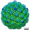
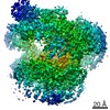
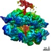
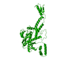
Journal: Elife / Year: 2020
Title: Cryo-EM structure in situ reveals a molecular switch that safeguards virus against genome loss.
Authors: Oliver W Bayfield / Alasdair C Steven / Alfred A Antson /
Abstract: The portal protein is a key component of many double-stranded DNA viruses, governing capsid assembly and genome packaging. Twelve subunits of the portal protein define a tunnel, through which DNA is ...The portal protein is a key component of many double-stranded DNA viruses, governing capsid assembly and genome packaging. Twelve subunits of the portal protein define a tunnel, through which DNA is translocated into the capsid. It is unknown how the portal protein functions as a gatekeeper, preventing DNA slippage, whilst allowing its passage into the capsid, and how these processes are controlled. A cryo-EM structure of the portal protein of thermostable virus P23-45, determined in situ in its procapsid-bound state, indicates a mechanism that naturally safeguards the virus against genome loss. This occurs via an inversion of the conformation of the loops that define the constriction in the central tunnel, accompanied by a hydrophilic-hydrophobic switch. The structure also shows how translocation of DNA into the capsid could be modulated by a changing mode of protein-protein interactions between portal and capsid, across a symmetry-mismatched interface.
History
DepositionJan 24, 2019Deposition site: PDBE / Processing site: PDBE
Revision 1.0Mar 25, 2020Provider: repository / Type: Initial release
Revision 1.1Apr 22, 2020Group: Database references / Category: citation / citation_author
Item: _citation.country / _citation.journal_abbrev ..._citation.country / _citation.journal_abbrev / _citation.journal_id_CSD / _citation.journal_id_ISSN / _citation.journal_volume / _citation.pdbx_database_id_DOI / _citation.pdbx_database_id_PubMed / _citation.title / _citation.year
Revision 1.2Jun 3, 2020Group: Database references / Category: citation / citation_author / Item: _citation.title / _citation_author.identifier_ORCID
Revision 1.3May 15, 2024Group: Data collection / Database references / Derived calculations
Category: chem_comp_atom / chem_comp_bond ...chem_comp_atom / chem_comp_bond / database_2 / pdbx_struct_oper_list
Item: _database_2.pdbx_DOI / _database_2.pdbx_database_accession ..._database_2.pdbx_DOI / _database_2.pdbx_database_accession / _pdbx_struct_oper_list.name / _pdbx_struct_oper_list.symmetry_operation

-
Structure visualization

Movie
  • Biological unit as author_defined_assembly
  • Imaged by Jmol
  • Download
  • Deposited structure unit
  • Imaged by Jmol
  • Download
  • Simplified surface model + fitted atomic model
  • EMDB-4567
  • Imaged by Jmol
  • Download
  • Superimposition on EM map
  • EMDB-4567
  • Imaged by UCSF Chimera
  • Download
Movie viewer
Structure viewerMolecule:
MolmilJmol/JSmol

Downloads & links

-
Assembly

Deposited unit
A: Portal protein


Theoretical massNumber of molelcules
Total (without water)49,7141
Polymers49,7141
Non-polymers00
Water0
1
A: Portal protein
x 12


Theoretical massNumber of molelcules
Total (without water)596,57012
Polymers596,57012
Non-polymers00
Water0
TypeNameSymmetry operationNumber
identity operation1_555x,y,z1
point symmetry operation11

-
Components

#1: Protein Portal protein


Mass: 49714.141 Da / Num. of mol.: 1 / Source method: isolated from a natural source / Source: (natural) Thermus virus P23-45 / Cell line: Thermus thermophilus HB8 / References: UniProt: A7XXB9

-
Experimental details

-
Experiment

ExperimentMethod: ELECTRON MICROSCOPY
EM experimentAggregation state: PARTICLE / 3D reconstruction method: single particle reconstruction

-
Sample preparation

ComponentName: Thermus phage P2345 / Type: COMPLEX / Entity ID: all / Source: NATURAL
Molecular weightValue: 0.596 MDa / Experimental value: YES
Source (natural)Organism: Thermus phage P2345 (virus)
Details of virusEmpty: YES / Enveloped: NO / Isolate: SPECIES / Type: VIRION
Natural hostOrganism: Thermus thermophilus HB8
Virus shellTriangulation number (T number): 7
Buffer solutionpH: 8
Buffer component
IDConc.FormulaBuffer-ID
1100 mMNaClSodium chloride1
220 mMTris-HClTris1
310 mMMgCl21
SpecimenEmbedding applied: NO / Shadowing applied: NO / Staining applied: NO / Vitrification applied: YES
VitrificationInstrument: FEI VITROBOT MARK IV / Cryogen name: ETHANE / Humidity: 90 % / Chamber temperature: 283 K

-
Electron microscopy imaging

Experimental equipment
Model: Titan Krios / Image courtesy: FEI Company
MicroscopyModel: FEI TITAN KRIOS
Electron gunElectron source: FIELD EMISSION GUN / Accelerating voltage: 300 kV / Illumination mode: FLOOD BEAM
Electron lensMode: BRIGHT FIELDBright-field microscopy / Nominal magnification: 75000 X / Nominal defocus max: 2500 nm / Nominal defocus min: 500 nm / Cs: 2.7 mm / C2 aperture diameter: 70 µm
Specimen holderCryogen: NITROGEN
Image recordingAverage exposure time: 2 sec. / Electron dose: 99 e/Å2 / Detector mode: INTEGRATING / Film or detector model: FEI FALCON III (4k x 4k)

-
Processing

SoftwareName: PHENIX / Version: 1.13_2998: / Classification: refinement
EM software
IDNameCategory
1RELIONparticle selection
2Scipionparticle selection
3EPUimage acquisition
5CTFFINDCTF correction
8Cootmodel fitting
10RELIONinitial Euler assignment
11RELIONfinal Euler assignment
12RELIONclassification
13RELION3D reconstruction
14PHENIXmodel refinement
15Cootmodel refinement
CTF correctionType: PHASE FLIPPING AND AMPLITUDE CORRECTION
SymmetryPoint symmetry: C12 (12 fold cyclic)
3D reconstructionResolution: 3.74 Å / Resolution method: FSC 0.143 CUT-OFF / Num. of particles: 10025 / Symmetry type: POINT
Atomic model buildingProtocol: AB INITIO MODEL / Space: REAL
RefinementHighest resolution: 3.74 Å
Refine LS restraints
Refine-IDTypeDev idealNumber
ELECTRON MICROSCOPYf_bond_d0.0053276
ELECTRON MICROSCOPYf_angle_d0.8424450
ELECTRON MICROSCOPYf_dihedral_angle_d11.7081964
ELECTRON MICROSCOPYf_chiral_restr0.051505
ELECTRON MICROSCOPYf_plane_restr0.007574

+
About Yorodumi

-
News

-
Feb 9, 2022. New format data for meta-information of EMDB entries

New format data for meta-information of EMDB entries

  • Version 3 of the EMDB header file is now the official format.
  • The previous official version 1.9 will be removed from the archive.

Related info.:EMDB header

External links:wwPDB to switch to version 3 of the EMDB data model

-
Aug 12, 2020. Covid-19 info

Covid-19 info

URL: https://pdbj.org/emnavi/covid19.php

New page: Covid-19 featured information page in EM Navigator.

Related info.:Covid-19 info / Mar 5, 2020. Novel coronavirus structure data

+
Mar 5, 2020. Novel coronavirus structure data

Novel coronavirus structure data

Related info.:Yorodumi Speices / Aug 12, 2020. Covid-19 info

External links:COVID-19 featured content - PDBj / Molecule of the Month (242):Coronavirus Proteases

+
Jan 31, 2019. EMDB accession codes are about to change! (news from PDBe EMDB page)

EMDB accession codes are about to change! (news from PDBe EMDB page)

  • The allocation of 4 digits for EMDB accession codes will soon come to an end. Whilst these codes will remain in use, new EMDB accession codes will include an additional digit and will expand incrementally as the available range of codes is exhausted. The current 4-digit format prefixed with “EMD-” (i.e. EMD-XXXX) will advance to a 5-digit format (i.e. EMD-XXXXX), and so on. It is currently estimated that the 4-digit codes will be depleted around Spring 2019, at which point the 5-digit format will come into force.
  • The EM Navigator/Yorodumi systems omit the EMD- prefix.

Related info.:Q: What is EMD? / ID/Accession-code notation in Yorodumi/EM Navigator

External links:EMDB Accession Codes are Changing Soon! / Contact to PDBj

+
Jul 12, 2017. Major update of PDB

Major update of PDB

  • wwPDB released updated PDB data conforming to the new PDBx/mmCIF dictionary.
  • This is a major update changing the version number from 4 to 5, and with Remediation, in which all the entries are updated.
  • In this update, many items about electron microscopy experimental information are reorganized (e.g. em_software).
  • Now, EM Navigator and Yorodumi are based on the updated data.

External links:wwPDB Remediation / Enriched Model Files Conforming to OneDep Data Standards Now Available in the PDB FTP Archive

-
Yorodumi

Thousand views of thousand structures

  • Yorodumi is a browser for structure data from EMDB, PDB, SASBDB, etc.
  • This page is also the successor to EM Navigator detail page, and also detail information page/front-end page for Omokage search.
  • The word "yorodu" (or yorozu) is an old Japanese word meaning "ten thousand". "mi" (miru) is to see.

Related info.:EMDB / PDB / SASBDB / Comparison of 3 databanks / Yorodumi Search / Aug 31, 2016. New EM Navigator & Yorodumi / Yorodumi Papers / Jmol/JSmol / Function and homology information / Changes in new EM Navigator and Yorodumi

Read more