 Movie
Movie Controller

[English] 日本語
 Yorodumi
- PDB-6q1f: Atomic structure of the Human Herpesvirus 6B Capsid and Capsid-As... -
+ Open data

- Basic information
| Entry | Database: PDB / ID: 6q1f | |||||||||||||||
|---|---|---|---|---|---|---|---|---|---|---|---|---|---|---|---|---|
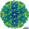
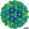
| Title | Atomic structure of the Human Herpesvirus 6B Capsid and Capsid-Associated Tegument Complexes | |||||||||||||||
 Components Components |
| |||||||||||||||
 Keywords Keywords | VIRUS / beta-herpesvirus / HHV-6B / murine cytomegalovirus / human cytomegalovirus / pp150 / pU11 / pUL32 / pM32 / pU14 | |||||||||||||||
| Function / homology |  Function and homology information Function and homology informationT=16 icosahedral viral capsid / viral tegument / viral capsid assembly / viral process / viral capsid / host cell nucleus / structural molecule activity / DNA binding Similarity search - Function | |||||||||||||||
| Biological species | Human herpesvirus 6B | |||||||||||||||
| Method | ELECTRON MICROSCOPY / single particle reconstruction / cryo EM / Resolution: 9 Å | |||||||||||||||
 Authors Authors | Zhang, Y.B. / Liu, W. / Li, Z.H. / Kumar, V. / Alvarez-Cabrera, A.L. / Leibovitch, E. / Cui, Y.X. / Mei, Y. / Bi, G.Q. / Jacobson, S. / Zhou, Z.H. | |||||||||||||||
| Funding support |  United States, 4items United States, 4items
| |||||||||||||||
 Citation Citation | Journal: Nat Commun / Year: 2019 Title: Atomic structure of the human herpesvirus 6B capsid and capsid-associated tegument complexes. Authors: Yibo Zhang / Wei Liu / Zihang Li / Vinay Kumar / Ana L Alvarez-Cabrera / Emily C Leibovitch / Yanxiang Cui / Ye Mei / Guo-Qiang Bi / Steve Jacobson / Z Hong Zhou /  Abstract: Human herpesvirus 6B (HHV-6B) belongs to the β-herpesvirus subfamily of the Herpesviridae. To understand capsid assembly and capsid-tegument interactions, here we report atomic structures of HHV-6B ...Human herpesvirus 6B (HHV-6B) belongs to the β-herpesvirus subfamily of the Herpesviridae. To understand capsid assembly and capsid-tegument interactions, here we report atomic structures of HHV-6B capsid and capsid-associated tegument complex (CATC) obtained by cryoEM and sub-particle reconstruction. Compared to other β-herpesviruses, HHV-6B exhibits high similarity in capsid structure but organizational differences in its CATC (pU11 tetramer). 180 "VΛ"-shaped CATCs are observed in HHV-6B, distinguishing from the 255 "Λ"-shaped dimeric CATCs observed in murine cytomegalovirus and the 310 "Δ"-shaped CATCs in human cytomegalovirus. This trend in CATC quantity correlates with the increasing genomes sizes of these β-herpesviruses. Incompatible distances revealed by the atomic structures rationalize the lack of CATC's binding to triplexes Ta, Tc, and Tf in HHV-6B. Our results offer insights into HHV-6B capsid assembly and the roles of its tegument proteins, including not only the β-herpesvirus-specific pU11 and pU14, but also those conserved across all subfamilies of Herpesviridae. | |||||||||||||||
| History |
|
- Structure visualization
| Movie |
Movie viewer |
|---|---|
| Structure viewer | Molecule: MolmilJmol/JSmol |
- Downloads & links
-Download
| PDBx/mmCIF format | 6q1f.cif.gz | 5.1 MB | Display | PDBx/mmCIF format |
|---|---|---|---|---|
| PDB format | pdb6q1f.ent.gz | Display | PDB format | |
| PDBx/mmJSON format | 6q1f.json.gz | Tree view | PDBx/mmJSON format | |
| Others |  Other downloads Other downloads |
-Validation report
| Summary document | 6q1f_validation.pdf.gz | 2 MB | Display | wwPDB validaton report |
|---|---|---|---|---|
| Full document | 6q1f_full_validation.pdf.gz | 2.1 MB | Display | |
| Data in XML | 6q1f_validation.xml.gz | 622.4 KB | Display | |
| Data in CIF | 6q1f_validation.cif.gz | 1004.1 KB | Display | |
| Arichive directory | https://data.pdbj.org/pub/pdb/validation_reports/q1/6q1fftp://data.pdbj.org/pub/pdb/validation_reports/q1/6q1f | HTTPS FTP |
-Related structure data
| Related structure data |  20557MC M: map data used to model this data C: citing same article ( |
|---|---|
| Similar structure data |

-Links
 PDBj
PDBj
- Assembly
| Deposited unit | 
|
|---|---|
| 1 | x 60
|
| 2 |
|
| 3 | x 5
|
| 4 | x 6
|
| 5 | 
|
| Symmetry | Point symmetry: (Schoenflies symbol: I (icosahedral)) |
-Components
| #1: Protein | Mass: 152260.047 Da / Num. of mol.: 16 / Source method: isolated from a natural source / Source: (natural)  Human herpesvirus 6B (strain Z29) / Strain: Z29 / References: UniProt: Q9QJ26 Human herpesvirus 6B (strain Z29) / Strain: Z29 / References: UniProt: Q9QJ26#2: Protein | Mass: 95738.125 Da / Num. of mol.: 12 / Source method: isolated from a natural source / Source: (natural) Human herpesvirus 6B (strain Z29) / Strain: Z29 / References: UniProt: Q69535#3: Protein | Mass: 9827.329 Da / Num. of mol.: 16 / Source method: isolated from a natural source / Source: (natural) Human herpesvirus 6B (strain Z29) / Strain: Z29 / References: UniProt: Q9WT32#4: Protein | Mass: 34162.508 Da / Num. of mol.: 5 / Source method: isolated from a natural source / Source: (natural) Human herpesvirus 6B (strain Z29) / Strain: Z29 / References: UniProt: Q9WT35#5: Protein | Mass: 33514.332 Da / Num. of mol.: 10 / Source method: isolated from a natural source / Source: (natural) Human herpesvirus 6B (strain Z29) / Strain: Z29 / References: UniProt: Q9QJ27 |
|---|
-Experimental details
-Experiment
| Experiment | Method: ELECTRON MICROSCOPY |
|---|---|
| EM experiment | Aggregation state: PARTICLE / 3D reconstruction method: single particle reconstruction |
- Sample preparation
| Component | Name: Human herpesvirus 6 strain Z29 / Type: VIRUS / Entity ID: all / Source: NATURAL |
|---|---|
| Molecular weight | Experimental value: NO |
| Source (natural) | Organism: Human herpesvirus 6 strain Z29 / Strain: Z29 |
| Details of virus | Empty: NO / Enveloped: YES / Isolate: STRAIN / Type: VIRION |
| Buffer solution | pH: 7.4 / Details: PBS buffer, pH 7.4 |
| Specimen | Embedding applied: NO / Shadowing applied: NO / Staining applied: NO / Vitrification applied: YES |
| Specimen support | Grid material: COPPER / Grid mesh size: 200 divisions/in. |
| Vitrification | Instrument: HOMEMADE PLUNGER / Cryogen name: ETHANE / Details: The grids were manually plunged into the ethane. |
- Electron microscopy imaging
| Experimental equipment |  Model: Titan Krios / Image courtesy: FEI Company |
|---|---|
| Microscopy | Model: FEI TITAN KRIOS |
| Electron gun | Electron source:  FIELD EMISSION GUN / Accelerating voltage: 300 kV / Illumination mode: FLOOD BEAM FIELD EMISSION GUN / Accelerating voltage: 300 kV / Illumination mode: FLOOD BEAM |
| Electron lens | Mode: BRIGHT FIELD / Nominal magnification: 64000 X / Calibrated magnification: 64000 X / Nominal defocus max: 3200 nm / Nominal defocus min: 2200 nm / Calibrated defocus min: 2200 nm / Calibrated defocus max: 3200 nm / Cs: 2.7 mm / Alignment procedure: COMA FREE |
| Specimen holder | Cryogen: NITROGEN / Specimen holder model: FEI TITAN KRIOS AUTOGRID HOLDER |
| Image recording | Electron dose: 23 e/Å2 / Film or detector model: GATAN K2 SUMMIT (4k x 4k) / Num. of real images: 4828 |
- Processing
| EM software |
| ||||||||||||
|---|---|---|---|---|---|---|---|---|---|---|---|---|---|
| CTF correction | Type: PHASE FLIPPING AND AMPLITUDE CORRECTION | ||||||||||||
| Particle selection | Num. of particles selected: 7430 | ||||||||||||
| Symmetry | Point symmetry: I (icosahedral) | ||||||||||||
| 3D reconstruction | Resolution: 9 Å / Resolution method: FSC 0.143 CUT-OFF / Num. of particles: 6443 / Details: bin4 reconstrction / Symmetry type: POINT | ||||||||||||
| Atomic model building | Protocol: OTHER |
