 Movie
Movie Controller

[English] 日本語
 Yorodumi
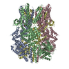
- PDB-6o6r: Structure of the TRPM8 cold receptor by single particle electron ... -
+ Open data

- Basic information
| Entry | Database: PDB / ID: 6o6r | |||||||||||||||
|---|---|---|---|---|---|---|---|---|---|---|---|---|---|---|---|---|
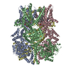
| Title | Structure of the TRPM8 cold receptor by single particle electron cryo-microscopy, AMTB-bound state | |||||||||||||||
 Components Components | Transient receptor potential cation channel subfamily M member 8 | |||||||||||||||
 Keywords Keywords | TRANSPORT PROTEIN / Ion Channel / TRPM8 | |||||||||||||||
| Function / homology | Chem-9PE / Chem-LQ7 / UNDECANE / CHOLESTEROL HEMISUCCINATE Function and homology information Function and homology information | |||||||||||||||
| Biological species |  Parus major (Great Tit) Parus major (Great Tit) | |||||||||||||||
| Method | ELECTRON MICROSCOPY / single particle reconstruction / cryo EM / Resolution: 3.2 Å | |||||||||||||||
 Authors Authors | Diver, M.M. / Cheng, Y. / Julius, D. | |||||||||||||||
| Funding support |  United States, 4items United States, 4items
| |||||||||||||||
 Citation Citation | Journal: Science / Year: 2019 Title: Structural insights into TRPM8 inhibition and desensitization. Authors: Melinda M Diver / Yifan Cheng / David Julius / Abstract: The transient receptor potential melastatin 8 (TRPM8) ion channel is the primary detector of environmental cold and an important target for treating pathological cold hypersensitivity. Here, we ...The transient receptor potential melastatin 8 (TRPM8) ion channel is the primary detector of environmental cold and an important target for treating pathological cold hypersensitivity. Here, we present cryo-electron microscopy structures of TRPM8 in ligand-free, antagonist-bound, or calcium-bound forms, revealing how robust conformational changes give rise to two nonconducting states, closed and desensitized. We describe a malleable ligand-binding pocket that accommodates drugs of diverse chemical structures, and we delineate the ion permeation pathway, including the contribution of lipids to pore architecture. Furthermore, we show that direct calcium binding mediates stimulus-evoked desensitization, clarifying this important mechanism of sensory adaptation. We observe large rearrangements within the S4-S5 linker that reposition the S1-S4 and pore domains relative to the TRP helix, leading us to propose a distinct model for modulation of TRPM8 and possibly other TRP channels. | |||||||||||||||
| History |
|
- Structure visualization
| Movie |
Movie viewer |
|---|---|
| Structure viewer | Molecule: MolmilJmol/JSmol |
- Downloads & links
-Download
| PDBx/mmCIF format | 6o6r.cif.gz | 624.5 KB | Display | PDBx/mmCIF format |
|---|---|---|---|---|
| PDB format | pdb6o6r.ent.gz | 502.2 KB | Display | PDB format |
| PDBx/mmJSON format | 6o6r.json.gz | Tree view | PDBx/mmJSON format | |
| Others |  Other downloads Other downloads |
-Validation report
| Arichive directory | https://data.pdbj.org/pub/pdb/validation_reports/o6/6o6rftp://data.pdbj.org/pub/pdb/validation_reports/o6/6o6r | HTTPS FTP |
|---|
-Related structure data
| Related structure data | 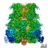 0636MC 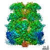 0631C 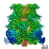 0638C  0639C  6o6aC  6o72C  6o77C M: map data used to model this data C: citing same article ( |
|---|---|
| Similar structure data |


-Links
 PDBj
PDBj
- Assembly
| Deposited unit | 
|
|---|---|
| 1 |
|
-Components
-Protein , 1 types, 4 molecules ABCD
| #1: Protein | Mass: 126989.797 Da / Num. of mol.: 4 Source method: isolated from a genetically manipulated source Source: (gene. exp.) Parus major (Great Tit) / Production host:  Homo sapiens (human) Homo sapiens (human) |
|---|
-Non-polymers , 5 types, 28 molecules 
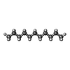
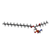
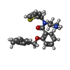

| #2: Chemical | ChemComp-Y01 /  Mass: 486.726 Da / Num. of mol.: 4 / Source method: obtained synthetically / Formula: C31H50O4 Mass: 486.726 Da / Num. of mol.: 4 / Source method: obtained synthetically / Formula: C31H50O4#3: Chemical | ChemComp-UND /  Mass: 156.308 Da / Num. of mol.: 8 / Source method: obtained synthetically / Formula: C11H24 Mass: 156.308 Da / Num. of mol.: 8 / Source method: obtained synthetically / Formula: C11H24#4: Chemical | ChemComp-9PE / (  Mass: 593.773 Da / Num. of mol.: 4 / Source method: obtained synthetically / Formula: C30H60NO8P / Comment: phospholipid*YM Mass: 593.773 Da / Num. of mol.: 4 / Source method: obtained synthetically / Formula: C30H60NO8P / Comment: phospholipid*YM#5: Chemical | ChemComp-LQ7 /  Mass: 394.530 Da / Num. of mol.: 4 / Source method: obtained synthetically / Formula: C23H26N2O2S Mass: 394.530 Da / Num. of mol.: 4 / Source method: obtained synthetically / Formula: C23H26N2O2S#6: Chemical | ChemComp-NA /  Mass: 22.990 Da / Num. of mol.: 8 / Source method: obtained synthetically / Formula: Na Mass: 22.990 Da / Num. of mol.: 8 / Source method: obtained synthetically / Formula: Na |
|---|
-Experimental details
-Experiment
| Experiment | Method: ELECTRON MICROSCOPY |
|---|---|
| EM experiment | Aggregation state: PARTICLE / 3D reconstruction method: single particle reconstruction |
- Sample preparation
| Component | Name: Transient receptor potential cation channel subfamily M member 8 Type: COMPLEX / Entity ID: #1 / Source: RECOMBINANT |
|---|---|
| Source (natural) | Organism: Parus major (Great Tit) |
| Source (recombinant) | Organism: Homo sapiens (human) |
| Buffer solution | pH: 7.4 |
| Specimen | Embedding applied: NO / Shadowing applied: NO / Staining applied: NO / Vitrification applied: YES |
| Specimen support | Details: unspecified |
| Vitrification | Cryogen name: ETHANE |
- Electron microscopy imaging
| Experimental equipment |  Model: Titan Krios / Image courtesy: FEI Company |
|---|---|
| Microscopy | Model: FEI TITAN KRIOS |
| Electron gun | Electron source:  FIELD EMISSION GUN / Accelerating voltage: 300 kV / Illumination mode: FLOOD BEAM FIELD EMISSION GUN / Accelerating voltage: 300 kV / Illumination mode: FLOOD BEAM |
| Electron lens | Mode: BRIGHT FIELD / Nominal magnification: 22500 X / Nominal defocus max: 2000 nm / Nominal defocus min: 600 nm |
| Image recording | Electron dose: 70 e/Å2 / Film or detector model: GATAN K2 SUMMIT (4k x 4k) |
- Processing
| CTF correction | Type: PHASE FLIPPING AND AMPLITUDE CORRECTION |
|---|---|
| Symmetry | Point symmetry: C4 (4 fold cyclic) |
| 3D reconstruction | Resolution: 3.2 Å / Resolution method: FSC 0.143 CUT-OFF / Num. of particles: 92836 / Symmetry type: POINT |
