 Movie
Movie Controller

[English] 日本語
 Yorodumi
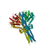
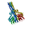
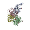
- PDB-6nt9: Cryo-EM structure of the complex between human TBK1 and chicken STING -
+ Open data

- Basic information
| Entry | Database: PDB / ID: 6nt9 | |||||||||||||||||||||
|---|---|---|---|---|---|---|---|---|---|---|---|---|---|---|---|---|---|---|---|---|---|---|
| Title | Cryo-EM structure of the complex between human TBK1 and chicken STING | |||||||||||||||||||||
 Components Components |
| |||||||||||||||||||||
 Keywords Keywords | IMMUNE SYSTEM / ER / kinase / adaptor | |||||||||||||||||||||
| Function / homology |  Function and homology information Function and homology informationSTING mediated induction of host immune responses / IRF3 mediated activation of type 1 IFN / positive regulation of xenophagy / STAT6-mediated induction of chemokines / dendritic cell proliferation / positive regulation of TORC2 signaling / serine/threonine protein kinase complex / regulation of type I interferon production / T follicular helper cell differentiation / 2',3'-cyclic GMP-AMP binding ...STING mediated induction of host immune responses / IRF3 mediated activation of type 1 IFN / positive regulation of xenophagy / STAT6-mediated induction of chemokines / dendritic cell proliferation / positive regulation of TORC2 signaling / serine/threonine protein kinase complex / regulation of type I interferon production / T follicular helper cell differentiation / 2',3'-cyclic GMP-AMP binding / cyclic-di-GMP binding / Interleukin-37 signaling / positive regulation of type I interferon-mediated signaling pathway / IRF3-mediated induction of type I IFN / proton channel activity / reticulophagy / TNFR1-induced proapoptotic signaling / TRAF6 mediated IRF7 activation / cGAS/STING signaling pathway / toll-like receptor 4 signaling pathway / peptidyl-threonine phosphorylation / type I interferon-mediated signaling pathway / cytoplasmic pattern recognition receptor signaling pathway / protein complex oligomerization / positive regulation of interferon-alpha production / autophagosome membrane / positive regulation of macroautophagy / autophagosome assembly / positive regulation of type I interferon production / canonical NF-kappaB signal transduction / negative regulation of TORC1 signaling / endoplasmic reticulum-Golgi intermediate compartment membrane / positive regulation of TORC1 signaling / activation of innate immune response / positive regulation of interferon-beta production / Regulation of TBK1, IKKε (IKBKE)-mediated activation of IRF3, IRF7 / positive regulation of autophagy / Regulation of TBK1, IKKε-mediated activation of IRF3, IRF7 upon TLR3 ligation / autophagosome / TICAM1-dependent activation of IRF3/IRF7 / Neutrophil degranulation / antiviral innate immune response / Regulation of innate immune responses to cytosolic DNA / peptidyl-serine phosphorylation / Activation of IRF3, IRF7 mediated by TBK1, IKKε (IKBKE) / PINK1-PRKN Mediated Mitophagy / macroautophagy / Negative regulators of DDX58/IFIH1 signaling / Regulation of TNFR1 signaling / phosphoprotein binding / DDX58/IFIH1-mediated induction of interferon-alpha/beta / response to virus / SARS-CoV-1 activates/modulates innate immune responses / TRAF3-dependent IRF activation pathway / cytoplasmic vesicle / protein phosphatase binding / Potential therapeutics for SARS / defense response to virus / nucleic acid binding / protein phosphorylation / RNA polymerase II-specific DNA-binding transcription factor binding / protein kinase activity / positive regulation of canonical NF-kappaB signal transduction / non-specific serine/threonine protein kinase / defense response to Gram-positive bacterium / inflammatory response / Golgi membrane / negative regulation of gene expression / innate immune response / protein serine kinase activity / protein serine/threonine kinase activity / endoplasmic reticulum membrane / SARS-CoV-2 activates/modulates innate and adaptive immune responses / perinuclear region of cytoplasm / protein homodimerization activity / positive regulation of transcription by RNA polymerase II / nucleoplasm / ATP binding / identical protein binding / cytoplasm / cytosol Similarity search - Function | |||||||||||||||||||||
| Biological species |  Homo sapiens (human) Homo sapiens (human) | |||||||||||||||||||||
| Method | ELECTRON MICROSCOPY / single particle reconstruction / cryo EM / Resolution: 3.3 Å | |||||||||||||||||||||
 Authors Authors | Shang, G. / Zhang, C. / Chen, Z.J. / Bai, X. / Zhang, X. | |||||||||||||||||||||
| Funding support |  United States, 6items United States, 6items
| |||||||||||||||||||||
 Citation Citation | Journal: Nature / Year: 2019 Title: Structural basis of STING binding with and phosphorylation by TBK1. Authors: Conggang Zhang / Guijun Shang / Xiang Gui / Xuewu Zhang / Xiao-Chen Bai / Zhijian J Chen / Abstract: The invasion of mammalian cytoplasm by microbial DNA from infectious pathogens or by self DNA from the nucleus or mitochondria represents a danger signal that alerts the host immune system. Cyclic ...The invasion of mammalian cytoplasm by microbial DNA from infectious pathogens or by self DNA from the nucleus or mitochondria represents a danger signal that alerts the host immune system. Cyclic GMP-AMP synthase (cGAS) is a sensor of cytoplasmic DNA that activates the type-I interferon pathway. On binding to DNA, cGAS is activated to catalyse the synthesis of cyclic GMP-AMP (cGAMP) from GTP and ATP. cGAMP functions as a second messenger that binds to and activates stimulator of interferon genes (STING). STING then recruits and activates tank-binding kinase 1 (TBK1), which phosphorylates STING and the transcription factor IRF3 to induce type-I interferons and other cytokines. However, how cGAMP-bound STING activates TBK1 and IRF3 is not understood. Here we present the cryo-electron microscopy structure of human TBK1 in complex with cGAMP-bound, full-length chicken STING. The structure reveals that the C-terminal tail of STING adopts a β-strand-like conformation and inserts into a groove between the kinase domain of one TBK1 subunit and the scaffold and dimerization domain of the second subunit in the TBK1 dimer. In this binding mode, the phosphorylation site Ser366 in the STING tail cannot reach the kinase-domain active site of bound TBK1, which suggests that STING phosphorylation by TBK1 requires the oligomerization of both proteins. Mutational analyses validate the interaction mode between TBK1 and STING and support a model in which high-order oligomerization of STING and TBK1, induced by cGAMP, leads to STING phosphorylation by TBK1. | |||||||||||||||||||||
| History |
|
- Structure visualization
| Movie |
Movie viewer |
|---|---|
| Structure viewer | Molecule: MolmilJmol/JSmol |
- Downloads & links
-Download
| PDBx/mmCIF format | 6nt9.cif.gz | 244.8 KB | Display | PDBx/mmCIF format |
|---|---|---|---|---|
| PDB format | pdb6nt9.ent.gz | 184.5 KB | Display | PDB format |
| PDBx/mmJSON format | 6nt9.json.gz | Tree view | PDBx/mmJSON format | |
| Others |  Other downloads Other downloads |
-Validation report
| Arichive directory | https://data.pdbj.org/pub/pdb/validation_reports/nt/6nt9ftp://data.pdbj.org/pub/pdb/validation_reports/nt/6nt9 | HTTPS FTP |
|---|
-Related structure data
| Related structure data |  0506MC M: map data used to model this data C: citing same article ( |
|---|---|
| Similar structure data |


-Links
 PDBj
PDBj


- Assembly
| Deposited unit | 
|
|---|---|
| 1 |
|
-Components
| #1: Protein | Mass: 85316.695 Da / Num. of mol.: 2 / Mutation: D135N Source method: isolated from a genetically manipulated source Source: (gene. exp.) Homo sapiens (human) / Gene: TBK1, NAK / Production host: Homo sapiens (human)References: UniProt: Q9UHD2, non-specific serine/threonine protein kinase #2: Protein | Mass: 44207.070 Da / Num. of mol.: 2 Source method: isolated from a genetically manipulated source Source: (gene. exp.) Homo sapiens (human) / References: UniProt: A0A1D5P7Q9, UniProt: E1C7U0*PLUS |
|---|
-Experimental details
-Experiment
| Experiment | Method: ELECTRON MICROSCOPY |
|---|---|
| EM experiment | Aggregation state: PARTICLE / 3D reconstruction method: single particle reconstruction |
- Sample preparation
| Component |
| ||||||||||||||||||||||||
|---|---|---|---|---|---|---|---|---|---|---|---|---|---|---|---|---|---|---|---|---|---|---|---|---|---|
| Source (natural) |
| ||||||||||||||||||||||||
| Source (recombinant) |
| ||||||||||||||||||||||||
| Buffer solution | pH: 8 | ||||||||||||||||||||||||
| Specimen | Conc.: 4.5 mg/ml / Embedding applied: NO / Shadowing applied: NO / Staining applied: NO / Vitrification applied: YES | ||||||||||||||||||||||||
| Specimen support | Details: unspecified | ||||||||||||||||||||||||
| Vitrification | Cryogen name: ETHANE |
- Electron microscopy imaging
| Experimental equipment |  Model: Titan Krios / Image courtesy: FEI Company |
|---|---|
| Microscopy | Model: FEI TITAN KRIOS |
| Electron gun | Electron source:  FIELD EMISSION GUN / Accelerating voltage: 300 kV / Illumination mode: FLOOD BEAM FIELD EMISSION GUN / Accelerating voltage: 300 kV / Illumination mode: FLOOD BEAM |
| Electron lens | Mode: BRIGHT FIELD |
| Specimen holder | Cryogen: NITROGEN / Specimen holder model: FEI TITAN KRIOS AUTOGRID HOLDER |
| Image recording | Electron dose: 48 e/Å2 / Detector mode: SUPER-RESOLUTION / Film or detector model: GATAN K2 SUMMIT (4k x 4k) |
| EM imaging optics | Energyfilter name: GIF Quantum LS / Energyfilter slit width: 20 eV |
| Image scans | Movie frames/image: 30 |
- Processing
| EM software |
| ||||||||||||||||||||||||||||||
|---|---|---|---|---|---|---|---|---|---|---|---|---|---|---|---|---|---|---|---|---|---|---|---|---|---|---|---|---|---|---|---|
| CTF correction | Type: PHASE FLIPPING AND AMPLITUDE CORRECTION | ||||||||||||||||||||||||||||||
| Symmetry | Point symmetry: C2 (2 fold cyclic) | ||||||||||||||||||||||||||||||
| 3D reconstruction | Resolution: 3.3 Å / Resolution method: FSC 0.143 CUT-OFF / Num. of particles: 86276 / Symmetry type: POINT | ||||||||||||||||||||||||||||||
| Atomic model building | Protocol: AB INITIO MODEL / Space: REAL | ||||||||||||||||||||||||||||||
| Atomic model building | PDB-ID: 4IM0 Accession code: 4IM0 / Source name: PDB / Type: experimental model |

