 Movie
Movie Controller


+ Open data

- Basic information
| Entry | Database: PDB / ID: 6ncl | ||||||
|---|---|---|---|---|---|---|---|
| Title | Near-atomic structure of icosahedrally averaged PBCV-1 capsid | ||||||
 Components Components |
| ||||||
 Keywords Keywords | VIRUS / tape-measure protein / minor capsid proteins / zip protein / giant virus | ||||||
| Function / homology |  Function and homology information Function and homology informationviral capsid / lipid binding / virion membrane / structural molecule activity Similarity search - Function | ||||||
| Biological species |  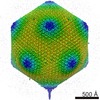 Paramecium bursaria Chlorella virus 1 Paramecium bursaria Chlorella virus 1 | ||||||
| Method | ELECTRON MICROSCOPY / single particle reconstruction / cryo EM / Resolution: 3.5 Å | ||||||
 Authors Authors | Fang, Q. / Rossmann, M.G. | ||||||
| Funding support |  United States, 1items United States, 1items
| ||||||
 Citation Citation | Journal: Nat Commun / Year: 2019 Title: Near-atomic structure of a giant virus. Authors: Qianglin Fang / Dongjie Zhu / Irina Agarkova / Jagat Adhikari / Thomas Klose / Yue Liu / Zhenguo Chen / Yingyuan Sun / Michael L Gross / James L Van Etten / Xinzheng Zhang / Michael G Rossmann /  Abstract: Although the nucleocytoplasmic large DNA viruses (NCLDVs) are one of the largest group of viruses that infect many eukaryotic hosts, the near-atomic resolution structures of these viruses have ...Although the nucleocytoplasmic large DNA viruses (NCLDVs) are one of the largest group of viruses that infect many eukaryotic hosts, the near-atomic resolution structures of these viruses have remained unknown. Here we describe a 3.5 Å resolution icosahedrally averaged capsid structure of Paramecium bursaria chlorella virus 1 (PBCV-1). This structure consists of 5040 copies of the major capsid protein, 60 copies of the penton protein and 1800 minor capsid proteins of which there are 13 different types. The minor capsid proteins form a hexagonal network below the outer capsid shell, stabilizing the capsid by binding neighboring capsomers together. The size of the viral capsid is determined by a tape-measure, minor capsid protein of which there are 60 copies in the virion. Homologs of the tape-measure protein and some of the other minor capsid proteins exist in other NCLDVs. Thus, a similar capsid assembly pathway might be used by other NCLDVs. | ||||||
| History |
|
- Structure visualization
| Movie |
Movie viewer |
|---|---|
| Structure viewer | Molecule: MolmilJmol/JSmol |
- Downloads & links
-Download
| PDBx/mmCIF format | 6ncl.cif.gz | 7 MB | Display | PDBx/mmCIF format |
|---|---|---|---|---|
| PDB format | pdb6ncl.ent.gz | Display | PDB format | |
| PDBx/mmJSON format | 6ncl.json.gz | Tree view | PDBx/mmJSON format | |
| Others |  Other downloads Other downloads |
-Validation report
| Arichive directory | https://data.pdbj.org/pub/pdb/validation_reports/nc/6nclftp://data.pdbj.org/pub/pdb/validation_reports/nc/6ncl | HTTPS FTP |
|---|
-Related structure data
| Related structure data | 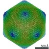 0436MC M: map data used to model this data C: citing same article ( |
|---|---|
| Similar structure data |
-Links
 PDBj
PDBj

- Assembly
| Deposited unit | 
|
|---|---|
| 1 | x 60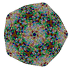
|
| 2 |
|
| 3 | x 5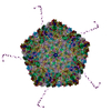
|
| 4 | x 6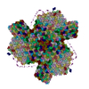
|
| 5 | 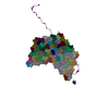
|
| Symmetry | Point symmetry: (Schoenflies symbol: I (icosahedral)) |
-Components
-Protein , 15 types, 115 molecules a0a1a2a3a4a5a6a7a8a9b0b1b2b3b4b5b7b8c0c1l5b6c2c3c4c5c6c7c8c9...
| #1: Protein | Mass: 38502.719 Da / Num. of mol.: 1 / Source method: isolated from a natural source / Source: (natural) Paramecium bursaria Chlorella virus 1 / References: UniProt: Q98550 | ||||||||||||||||||||||||
|---|---|---|---|---|---|---|---|---|---|---|---|---|---|---|---|---|---|---|---|---|---|---|---|---|---|
| #2: Protein | Mass: 23407.564 Da / Num. of mol.: 1 / Source method: isolated from a natural source / Source: (natural) Paramecium bursaria Chlorella virus 1 / References: UniProt: Q98459 | ||||||||||||||||||||||||
| #3: Protein | Mass: 31223.600 Da / Num. of mol.: 2 / Source method: isolated from a natural source / Source: (natural) Paramecium bursaria Chlorella virus 1 / References: UniProt: Q98505#4: Protein | | Mass: 29505.855 Da / Num. of mol.: 1 / Source method: isolated from a natural source / Source: (natural) Paramecium bursaria Chlorella virus 1 / References: UniProt: Q84580#5: Protein | | Mass: 24036.729 Da / Num. of mol.: 1 / Source method: isolated from a natural source / Source: (natural) Paramecium bursaria Chlorella virus 1 / References: UniProt: Q84523#6: Protein | | Mass: 18281.664 Da / Num. of mol.: 1 / Source method: isolated from a natural source / Source: (natural) Paramecium bursaria Chlorella virus 1 / References: UniProt: Q84626#7: Protein | | Mass: 17715.475 Da / Num. of mol.: 1 / Source method: isolated from a natural source / Source: (natural) Paramecium bursaria Chlorella virus 1 / References: UniProt: Q84459#8: Protein | | Mass: 16455.834 Da / Num. of mol.: 1 / Source method: isolated from a natural source / Source: (natural) Paramecium bursaria Chlorella virus 1 / References: UniProt: Q98576#9: Protein | Mass: 23322.656 Da / Num. of mol.: 12 / Source method: isolated from a natural source / Source: (natural) Paramecium bursaria Chlorella virus 1 / References: UniProt: Q84666#10: Protein | | Mass: 63888.082 Da / Num. of mol.: 1 / Source method: isolated from a natural source / Source: (natural) Paramecium bursaria Chlorella virus 1 / References: UniProt: Q84656#11: Protein | Mass: 20637.311 Da / Num. of mol.: 4 / Source method: isolated from a natural source / Source: (natural) Paramecium bursaria Chlorella virus 1 / References: UniProt: O41054#12: Protein | Mass: 19116.688 Da / Num. of mol.: 3 / Source method: isolated from a natural source / Source: (natural) Paramecium bursaria Chlorella virus 1 / References: UniProt: Q98573#13: Protein | | Mass: 19226.947 Da / Num. of mol.: 1 / Source method: isolated from a natural source / Source: (natural) Paramecium bursaria Chlorella virus 1 / References: UniProt: O41126#14: Protein | Mass: 48199.625 Da / Num. of mol.: 84 / Source method: isolated from a natural source / Source: (natural) Paramecium bursaria Chlorella virus 1 / References: UniProt: P30328#15: Protein | | Mass: 11070.830 Da / Num. of mol.: 1 / Source method: isolated from a natural source / Source: (natural) Paramecium bursaria Chlorella virus 1 / References: UniProt: Q98473 |
-Details
| Has protein modification | Y |
|---|
-Experimental details
-Experiment
| Experiment | Method: ELECTRON MICROSCOPY |
|---|---|
| EM experiment | Aggregation state: PARTICLE / 3D reconstruction method: single particle reconstruction |
- Sample preparation
| Component | Name: Paramecium bursaria Chlorella virus 1 / Type: VIRUS / Entity ID: all / Source: NATURAL |
|---|---|
| Source (natural) | Organism: Paramecium bursaria Chlorella virus 1 |
| Details of virus | Empty: NO / Enveloped: NO / Isolate: OTHER / Type: VIRION |
| Buffer solution | pH: 7.8 |
| Specimen | Embedding applied: NO / Shadowing applied: NO / Staining applied: NO / Vitrification applied: YES |
| Specimen support | Details: unspecified |
| Vitrification | Instrument: GATAN CRYOPLUNGE 3 / Cryogen name: ETHANE / Humidity: 85 % |
- Electron microscopy imaging
| Experimental equipment |  Model: Titan Krios / Image courtesy: FEI Company |
|---|---|
| Microscopy | Model: FEI TITAN KRIOS |
| Electron gun | Electron source:  FIELD EMISSION GUN / Accelerating voltage: 300 kV / Illumination mode: FLOOD BEAM FIELD EMISSION GUN / Accelerating voltage: 300 kV / Illumination mode: FLOOD BEAM |
| Electron lens | Mode: BRIGHT FIELD |
| Image recording | Electron dose: 24.4 e/Å2 / Detector mode: SUPER-RESOLUTION / Film or detector model: GATAN K2 SUMMIT (4k x 4k) |
- Processing
| CTF correction | Type: PHASE FLIPPING AND AMPLITUDE CORRECTION |
|---|---|
| Symmetry | Point symmetry: I (icosahedral) |
| 3D reconstruction | Resolution: 3.5 Å / Resolution method: FSC 0.143 CUT-OFF / Num. of particles: 13000 / Symmetry type: POINT |
