National Natural Science Foundation of China (NSFC)
31873012
中国
引用
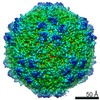
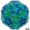
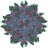
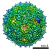
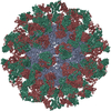
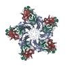
ジャーナル: J Virol / 年: 2020 タイトル: Neutralization Mechanism of a Monoclonal Antibody Targeting a Porcine Circovirus Type 2 Cap Protein Conformational Epitope. 著者: Liping Huang / Zhenzhao Sun / Deli Xia / Yanwu Wei / Encheng Sun / Chunguo Liu / Hongzhen Zhu / Haiqiao Bian / Hongli Wu / Li Feng / Jingfei Wang / Changming Liu / 要旨: Porcine circovirus type 2 (PCV2) is an important pathogen in swine herds, and its infection of pigs has caused severe economic losses to the pig industry worldwide. The capsid protein of PCV2 is the ...Porcine circovirus type 2 (PCV2) is an important pathogen in swine herds, and its infection of pigs has caused severe economic losses to the pig industry worldwide. The capsid protein of PCV2 is the only structural protein that is associated with PCV2 infection and immunity. Here, we report a neutralizing monoclonal antibody (MAb), MAb 3A5, that binds to intact PCV2 virions of the PCV2a, PCV2b, and PCV2d genotypes. MAb 3A5 neutralized PCV2 by blocking viral attachment to PK15 cells. To further explore the neutralization mechanism, we resolved the structure of the PCV2 virion in complex with MAb 3A5 Fab fragments by using cryo-electron microscopy single-particle analysis. The binding sites were located at the topmost edges around 5-fold icosahedral symmetry axes, with each footprint covering amino acids from two adjacent capsid proteins. Most of the epitope residues (15/18 residues) were conserved among 2,273 PCV2 strains. Mutations of some amino acids within the epitope had significant effects on the neutralizing activity of MAb 3A5. This study reveals the molecular and structural bases of this PCV2-neutralizing antibody and provides new and important information for vaccine design and therapeutic antibody development against PCV2 infections. PCV2 is associated with several clinical manifestations collectively known as PCV2-associated diseases (PCVADs). Neutralizing antibodies play a crucial role in the prevention of PCVADs. We demonstrated previously that a MAb, MAb 3A5, neutralizes the PCV2a, PCV2b, and PCV2d genotypes with different degrees of efficiency, but the underlying mechanism remains elusive. Here, we report the neutralization mechanism of this MAb and the structure of the PCV2 virion in complex with MAb 3A5 Fabs, showing a binding mode in which one Fab interacted with more than two loops from two adjacent capsid proteins. This binding mode has not been observed previously for PCV2-neutralizing antibodies. Our work provides new and important information for vaccine design and therapeutic antibody development against PCV2 infections.
 ムービー
ムービー コントローラー


 データを開く
 基本情報
基本情報 要素
要素 キーワード
キーワード 機能・相同性情報

 Porcine circovirus 2 (ウイルス)
 データ登録者
 中国, 1件
  引用
引用 構造の表示
構造の表示 ダウンロードとリンク
 その他のダウンロード


 PDBj
PDBj


 集合体
集合体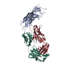


 試料調製
試料調製 電子顕微鏡撮影
 FIELD EMISSION GUN / 加速電圧: 200 kV / 照射モード: FLOOD BEAM
 解析
解析