 Movie
Movie Controller

[English] 日本語
 Yorodumi
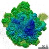
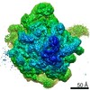
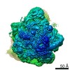
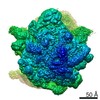
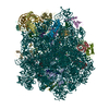
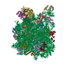
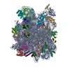
- PDB-6hma: Improved model derived from cryo-EM map of Staphylococcus aureus ... -
+ Open data

- Basic information
| Entry | Database: PDB / ID: 6hma | ||||||||||||||||||||||||||||||||||||||||||||||||||||||
|---|---|---|---|---|---|---|---|---|---|---|---|---|---|---|---|---|---|---|---|---|---|---|---|---|---|---|---|---|---|---|---|---|---|---|---|---|---|---|---|---|---|---|---|---|---|---|---|---|---|---|---|---|---|---|---|
| Title | Improved model derived from cryo-EM map of Staphylococcus aureus large ribosomal subunit | ||||||||||||||||||||||||||||||||||||||||||||||||||||||
 Components Components |
| ||||||||||||||||||||||||||||||||||||||||||||||||||||||
 Keywords Keywords | RIBOSOME / ribosomal RNA / pathogen / ribosomal protein / cryo-EM | ||||||||||||||||||||||||||||||||||||||||||||||||||||||
| Function / homology |  Function and homology information Function and homology informationlarge ribosomal subunit / transferase activity / 5S rRNA binding / ribosomal large subunit assembly / large ribosomal subunit rRNA binding / cytosolic large ribosomal subunit / cytoplasmic translation / tRNA binding / negative regulation of translation / rRNA binding ...large ribosomal subunit / transferase activity / 5S rRNA binding / ribosomal large subunit assembly / large ribosomal subunit rRNA binding / cytosolic large ribosomal subunit / cytoplasmic translation / tRNA binding / negative regulation of translation / rRNA binding / structural constituent of ribosome / ribosome / translation / ribonucleoprotein complex / mRNA binding / RNA binding / cytoplasm Similarity search - Function | ||||||||||||||||||||||||||||||||||||||||||||||||||||||
| Biological species |   Staphylococcus aureus (bacteria) Staphylococcus aureus (bacteria) | ||||||||||||||||||||||||||||||||||||||||||||||||||||||
| Method | ELECTRON MICROSCOPY / single particle reconstruction / cryo EM / Resolution: 2.65 Å | ||||||||||||||||||||||||||||||||||||||||||||||||||||||
 Authors Authors | Eyal, Z. / Cimicata, G. / Matzov, D. / Fox, T. / de Val, N. / Zimmerman, E. / Bashan, A. / Yonath, A. | ||||||||||||||||||||||||||||||||||||||||||||||||||||||
 Citation Citation | Journal: mBio / Year: 2022 Title: Structural Studies Reveal the Role of Helix 68 in the Elongation Step of Protein Biosynthesis. Authors: Giuseppe Cimicata / Gil Fridkin / Tanaya Bose / Zohar Eyal / Yehuda Halfon / Elinor Breiner-Goldstein / Tara Fox / Ella Zimmerman / Anat Bashan / Natalia de Val / Alexander Wlodawer / Ada Yonath /   Abstract: The ribosome, a multicomponent assembly consisting of RNA and proteins, is a pivotal macromolecular machine that translates the genetic code into proteins. The large ribosomal subunit rRNA helix 68 ...The ribosome, a multicomponent assembly consisting of RNA and proteins, is a pivotal macromolecular machine that translates the genetic code into proteins. The large ribosomal subunit rRNA helix 68 (H68) is a key element in the protein synthesis process, as it coordinates the coupled movements of the actors involved in translocation, including the tRNAs and L1 stalk. Examination of cryo-electron microscopy (cryo-EM) structures of ribosomes incubated for various time durations at physiological temperatures led to the identification of functionally relevant H68 movements. These movements assist the transition of the L1 stalk between its open and closed states. H68 spatial flexibility and its significance to the protein synthesis process were confirmed through its effective targeting with antisense PNA oligomers. Our results suggest that H68 is actively involved in ribosome movements that are central to the elongation process. The mechanism that regulates the translocation step in ribosomes during protein synthesis is not fully understood. In this work, cryo-EM techniques used to image ribosomes from Staphylococcus aureus after incubation at physiological temperature allowed the identification of a conformation of the helix 68 that has never been observed so far. We then propose a mechanism in which such helix, switching between two different conformations, actively coordinates the translocation step, shedding light on the dynamics of ribosomal components. In addition, the relevance of helix 68 to ribosome function and its potential as an antibiotic target was proved by inhibiting Staphylococcus aureus ribosomes activity using oligomers with sequence complementarity. #1: Journal: Proc Natl Acad Sci U S A / Year: 2015Title: Structural insights into species-specific features of the ribosome from the pathogen Staphylococcus aureus. Authors: Zohar Eyal / Donna Matzov / Miri Krupkin / Itai Wekselman / Susanne Paukner / Ella Zimmerman / Haim Rozenberg / Anat Bashan / Ada Yonath /  Abstract: The emergence of bacterial multidrug resistance to antibiotics threatens to cause regression to the preantibiotic era. Here we present the crystal structure of the large ribosomal subunit from ...The emergence of bacterial multidrug resistance to antibiotics threatens to cause regression to the preantibiotic era. Here we present the crystal structure of the large ribosomal subunit from Staphylococcus aureus, a versatile Gram-positive aggressive pathogen, and its complexes with the known antibiotics linezolid and telithromycin, as well as with a new, highly potent pleuromutilin derivative, BC-3205. These crystal structures shed light on specific structural motifs of the S. aureus ribosome and the binding modes of the aforementioned antibiotics. Moreover, by analyzing the ribosome structure and comparing it with those of nonpathogenic bacterial models, we identified some unique internal and peripheral structural motifs that may be potential candidates for improving known antibiotics and for use in the design of selective antibiotic drugs against S. aureus. | ||||||||||||||||||||||||||||||||||||||||||||||||||||||
| History |
|
- Structure visualization
| Movie |
Movie viewer |
|---|---|
| Structure viewer | Molecule: MolmilJmol/JSmol |
- Downloads & links
-Download
| PDBx/mmCIF format | 6hma.cif.gz | 1.9 MB | Display | PDBx/mmCIF format |
|---|---|---|---|---|
| PDB format | pdb6hma.ent.gz | 1.5 MB | Display | PDB format |
| PDBx/mmJSON format | 6hma.json.gz | Tree view | PDBx/mmJSON format | |
| Others |  Other downloads Other downloads |
-Validation report
| Arichive directory | https://data.pdbj.org/pub/pdb/validation_reports/hm/6hmaftp://data.pdbj.org/pub/pdb/validation_reports/hm/6hma | HTTPS FTP |
|---|
-Related structure data
| Related structure data | 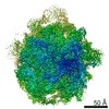 0243MC 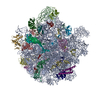 7asmC 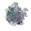 7asnC 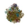 7asoC  7aspC M: map data used to model this data C: citing same article ( |
|---|---|
| Similar structure data |


-Links
 PDBj
PDBj


- Assembly
| Deposited unit | 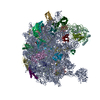
|
|---|---|
| 1 |
|
-Components
-RNA chain , 2 types, 2 molecules AB
| #1: RNA chain | Mass: 946738.688 Da / Num. of mol.: 1 / Source method: isolated from a natural source / Source: (natural) Staphylococcus aureus (bacteria) / References: GenBank: 1418434134 |
|---|---|
| #2: RNA chain | Mass: 36974.945 Da / Num. of mol.: 1 / Source method: isolated from a natural source / Source: (natural) Staphylococcus aureus (bacteria) / References: GenBank: 1434110260 |
+50S ribosomal protein ... , 27 types, 27 molecules CDEFGHIJKLMNOPQRSTUVWXZ1234
-Non-polymers , 1 types, 237 molecules 
| #30: Chemical | ChemComp-MG /  Mass: 24.305 Da / Num. of mol.: 237 / Source method: obtained synthetically / Formula: Mg Mass: 24.305 Da / Num. of mol.: 237 / Source method: obtained synthetically / Formula: Mg |
|---|
-Details
| Has protein modification | Y |
|---|
-Experimental details
-Experiment
| Experiment | Method: ELECTRON MICROSCOPY |
|---|---|
| EM experiment | Aggregation state: PARTICLE / 3D reconstruction method: single particle reconstruction |
- Sample preparation
| Component | Name: Staphylococcus aureus large ribosomal subunit / Type: RIBOSOME / Entity ID: #1-#29 / Source: NATURAL |
|---|---|
| Source (natural) | Organism: Staphylococcus aureus (bacteria) |
| Buffer solution | pH: 7.6 |
| Specimen | Conc.: 1 mg/ml / Embedding applied: NO / Shadowing applied: NO / Staining applied: NO / Vitrification applied: YES |
| Vitrification | Cryogen name: ETHANE |
- Electron microscopy imaging
| Experimental equipment |  Model: Titan Krios / Image courtesy: FEI Company |
|---|---|
| Microscopy | Model: FEI TITAN KRIOS |
| Electron gun | Electron source:  FIELD EMISSION GUN / Accelerating voltage: 300 kV / Illumination mode: FLOOD BEAM FIELD EMISSION GUN / Accelerating voltage: 300 kV / Illumination mode: FLOOD BEAM |
| Electron lens | Mode: BRIGHT FIELD |
| Image recording | Electron dose: 40 e/Å2 / Film or detector model: GATAN K2 SUMMIT (4k x 4k) |
- Processing
| Software | Name: PHENIX / Version: 1.13_2998: / Classification: refinement |
|---|---|
| CTF correction | Type: NONE |
| 3D reconstruction | Resolution: 2.65 Å / Resolution method: FSC 0.143 CUT-OFF / Num. of particles: 211046 / Symmetry type: POINT |
