 Movie
Movie Controller


+ Open data

- Basic information
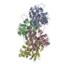
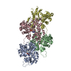
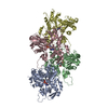
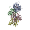
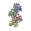
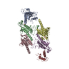
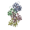
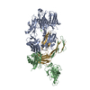
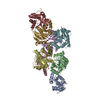
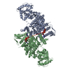
| Entry | Database: PDB / ID: 6djo | |||||||||
|---|---|---|---|---|---|---|---|---|---|---|
| Title | Cryo-EM structure of ADP-actin filaments | |||||||||
 Components Components | Actin, alpha skeletal muscle | |||||||||
 Keywords Keywords | CONTRACTILE PROTEIN / actin / ADP / filament / ATPase | |||||||||
| Function / homology |  Function and homology information Function and homology informationRegulation of CDH1 Function / Striated Muscle Contraction / striated muscle thin filament / skeletal muscle thin filament assembly / skeletal muscle fiber development / stress fiber / actin filament / Hydrolases; Acting on acid anhydrides; Acting on acid anhydrides to facilitate cellular and subcellular movement / actin cytoskeleton / hydrolase activity / ATP binding Similarity search - Function | |||||||||
| Biological species |  | |||||||||
| Method | ELECTRON MICROSCOPY / helical reconstruction / cryo EM / Resolution: 3.6 Å | |||||||||
 Authors Authors | Chou, S.Z. / Pollard, T.D. | |||||||||
| Funding support |  United States, 2items United States, 2items
| |||||||||
 Citation Citation | Journal: Proc Natl Acad Sci U S A / Year: 2019 Title: Mechanism of actin polymerization revealed by cryo-EM structures of actin filaments with three different bound nucleotides. Authors: Steven Z Chou / Thomas D Pollard / Abstract: We used cryo-electron microscopy (cryo-EM) to reconstruct actin filaments with bound AMPPNP (β,γ-imidoadenosine 5'-triphosphate, an ATP analog, resolution 3.1 Å), ADP-P (ADP with inorganic ...We used cryo-electron microscopy (cryo-EM) to reconstruct actin filaments with bound AMPPNP (β,γ-imidoadenosine 5'-triphosphate, an ATP analog, resolution 3.1 Å), ADP-P (ADP with inorganic phosphate, resolution 3.1 Å), or ADP (resolution 3.6 Å). Subunits in the three filaments have similar backbone conformations, so assembly rather than ATP hydrolysis or phosphate dissociation is responsible for their flattened conformation in filaments. Polymerization increases the rate of ATP hydrolysis by changing the positions of the side chains of Q137 and H161 in the active site. Flattening during assembly also promotes interactions along both the long-pitch and short-pitch helices. In particular, conformational changes in subdomain 3 open up multiple favorable interactions with the DNase-I binding loop in subdomain 2 of the adjacent subunit. Subunits at the barbed end of the filament are likely to be in this favorable conformation, while monomers are not. This difference explains why filaments grow faster at the barbed end than the pointed end. When phosphate dissociates from ADP-P-actin through a backdoor channel, the conformation of the C terminus changes so it distorts the DNase binding loop, which allows cofilin binding, and a network of interactions among S14, H73, G74, N111, R177, and G158 rearranges to open the phosphate release site. | |||||||||
| History |
|
- Structure visualization
| Movie |
Movie viewer |
|---|---|
| Structure viewer | Molecule: MolmilJmol/JSmol |
- Downloads & links
-Download
| PDBx/mmCIF format | 6djo.cif.gz | 261.4 KB | Display | PDBx/mmCIF format |
|---|---|---|---|---|
| PDB format | pdb6djo.ent.gz | 213.7 KB | Display | PDB format |
| PDBx/mmJSON format | 6djo.json.gz | Tree view | PDBx/mmJSON format | |
| Others |  Other downloads Other downloads |
-Validation report
| Arichive directory | https://data.pdbj.org/pub/pdb/validation_reports/dj/6djoftp://data.pdbj.org/pub/pdb/validation_reports/dj/6djo | HTTPS FTP |
|---|
-Related structure data
| Related structure data | 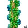 7938MC 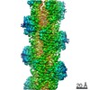 7936C 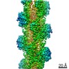 7937C 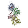 6djmC 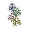 6djnC M: map data used to model this data C: citing same article ( |
|---|---|
| Similar structure data |
-Links
 PDBj
PDBj


- Assembly
| Deposited unit | 
|
|---|---|
| 1 |
|
-Components
| #1: Protein | Mass: 41875.633 Da / Num. of mol.: 4 / Source method: isolated from a natural source / Source: (natural) #2: Chemical | ChemComp-MG /   Mass: 24.305 Da / Num. of mol.: 4 / Source method: obtained synthetically / Formula: Mg Mass: 24.305 Da / Num. of mol.: 4 / Source method: obtained synthetically / Formula: Mg#3: Chemical | ChemComp-ADP /   Mass: 427.201 Da / Num. of mol.: 4 / Source method: obtained synthetically / Formula: C10H15N5O10P2 / Comment: ADP, energy-carrying molecule*YM Mass: 427.201 Da / Num. of mol.: 4 / Source method: obtained synthetically / Formula: C10H15N5O10P2 / Comment: ADP, energy-carrying molecule*YM |
|---|
-Experimental details
-Experiment
| Experiment | Method: ELECTRON MICROSCOPY |
|---|---|
| EM experiment | Aggregation state: FILAMENT / 3D reconstruction method: helical reconstruction |
- Sample preparation
| Component | Name: ADP-actin / Type: COMPLEX / Entity ID: #1 / Source: NATURAL |
|---|---|
| Source (natural) | Organism: |
| Buffer solution | pH: 7 |
| Specimen | Embedding applied: NO / Shadowing applied: NO / Staining applied: NO / Vitrification applied: YES |
| Specimen support | Details: unspecified |
| Vitrification | Cryogen name: ETHANE |
- Electron microscopy imaging
| Experimental equipment |  Model: Titan Krios / Image courtesy: FEI Company |
|---|---|
| Microscopy | Model: FEI TITAN KRIOS |
| Electron gun | Electron source:  FIELD EMISSION GUN / Accelerating voltage: 300 kV / Illumination mode: FLOOD BEAM FIELD EMISSION GUN / Accelerating voltage: 300 kV / Illumination mode: FLOOD BEAM |
| Electron lens | Mode: BRIGHT FIELD / Cs: 2.7 mm |
| Specimen holder | Cryogen: NITROGEN |
| Image recording | Electron dose: 43.54 e/Å2 / Detector mode: SUPER-RESOLUTION / Film or detector model: GATAN K2 SUMMIT (4k x 4k) |
| EM imaging optics | Energyfilter name: GIF Quantum LS / Energyfilter slit width: 20 eV |
- Processing
| Software | Name: PHENIX / Version: 1.12_2829: / Classification: refinement | ||||||||||||||||||||||||
|---|---|---|---|---|---|---|---|---|---|---|---|---|---|---|---|---|---|---|---|---|---|---|---|---|---|
| EM software |
| ||||||||||||||||||||||||
| CTF correction | Type: PHASE FLIPPING AND AMPLITUDE CORRECTION | ||||||||||||||||||||||||
| Helical symmerty | Angular rotation/subunit: -166.55 ° / Axial rise/subunit: 27.45 Å / Axial symmetry: C1 | ||||||||||||||||||||||||
| 3D reconstruction | Resolution: 3.6 Å / Resolution method: FSC 0.143 CUT-OFF / Num. of particles: 123806 / Symmetry type: HELICAL | ||||||||||||||||||||||||
| Atomic model building | Protocol: AB INITIO MODEL | ||||||||||||||||||||||||
| Refine LS restraints |
|
