 Movie
Movie Controller

[English] 日本語
 Yorodumi
- PDB-6c54: Ebola nucleoprotein nucleocapsid-like assembly and the asymmetric unit -
+ Open data

- Basic information
| Entry | Database: PDB / ID: 6c54 | ||||||||||||||||||||||||
|---|---|---|---|---|---|---|---|---|---|---|---|---|---|---|---|---|---|---|---|---|---|---|---|---|---|
| Title | Ebola nucleoprotein nucleocapsid-like assembly and the asymmetric unit | ||||||||||||||||||||||||
 Components Components | Nucleoprotein | ||||||||||||||||||||||||
 Keywords Keywords | RNA BINDING PROTEIN / Ebola / nucleoprotein / nucleocapsid / helical reconstruction | ||||||||||||||||||||||||
| Function / homology |  Function and homology information Function and homology informationviral RNA genome packaging / helical viral capsid / viral nucleocapsid / host cell cytoplasm / ribonucleoprotein complex / RNA binding Similarity search - Function | ||||||||||||||||||||||||
| Biological species |   Zaire ebolavirus Zaire ebolavirus | ||||||||||||||||||||||||
| Method | ELECTRON MICROSCOPY / helical reconstruction / cryo EM / Resolution: 5.8 Å | ||||||||||||||||||||||||
 Authors Authors | Su, Z. / Wu, C. / Pintilie, G.D. / Chiu, W. / Amarasinghe, G.K. / Leung, D.W. | ||||||||||||||||||||||||
| Funding support |  United States, 7items United States, 7items
| ||||||||||||||||||||||||
 Citation Citation | Journal: Cell / Year: 2018 Title: Electron Cryo-microscopy Structure of Ebola Virus Nucleoprotein Reveals a Mechanism for Nucleocapsid-like Assembly. Authors: Zhaoming Su / Chao Wu / Liuqing Shi / Priya Luthra / Grigore D Pintilie / Britney Johnson / Justin R Porter / Peng Ge / Muyuan Chen / Gai Liu / Thomas E Frederick / Jennifer M Binning / ...Authors: Zhaoming Su / Chao Wu / Liuqing Shi / Priya Luthra / Grigore D Pintilie / Britney Johnson / Justin R Porter / Peng Ge / Muyuan Chen / Gai Liu / Thomas E Frederick / Jennifer M Binning / Gregory R Bowman / Z Hong Zhou / Christopher F Basler / Michael L Gross / Daisy W Leung / Wah Chiu / Gaya K Amarasinghe / Abstract: Ebola virus nucleoprotein (eNP) assembles into higher-ordered structures that form the viral nucleocapsid (NC) and serve as the scaffold for viral RNA synthesis. However, molecular insights into the ...Ebola virus nucleoprotein (eNP) assembles into higher-ordered structures that form the viral nucleocapsid (NC) and serve as the scaffold for viral RNA synthesis. However, molecular insights into the NC assembly process are lacking. Using a hybrid approach, we characterized the NC-like assembly of eNP, identified novel regulatory elements, and described how these elements impact function. We generated a three-dimensional structure of the eNP NC-like assembly at 5.8 Å using electron cryo-microscopy and identified a new regulatory role for eNP helices α22-α23. Biochemical, biophysical, and mutational analyses revealed that inter-eNP contacts within α22-α23 are critical for viral NC assembly and regulate viral RNA synthesis. These observations suggest that the N terminus and α22-α23 of eNP function as context-dependent regulatory modules (CDRMs). Our current study provides a framework for a structural mechanism for NC-like assembly and a new therapeutic target. #1: Journal: Cell Rep / Year: 2015Title: An Intrinsically Disordered Peptide from Ebola Virus VP35 Controls Viral RNA Synthesis by Modulating Nucleoprotein-RNA Interactions. Authors: Daisy W Leung / Dominika Borek / Priya Luthra / Jennifer M Binning / Manu Anantpadma / Gai Liu / Ian B Harvey / Zhaoming Su / Ariel Endlich-Frazier / Juanli Pan / Reed S Shabman / Wah Chiu / ...Authors: Daisy W Leung / Dominika Borek / Priya Luthra / Jennifer M Binning / Manu Anantpadma / Gai Liu / Ian B Harvey / Zhaoming Su / Ariel Endlich-Frazier / Juanli Pan / Reed S Shabman / Wah Chiu / Robert A Davey / Zbyszek Otwinowski / Christopher F Basler / Gaya K Amarasinghe / Abstract: During viral RNA synthesis, Ebola virus (EBOV) nucleoprotein (NP) alternates between an RNA-template-bound form and a template-free form to provide the viral polymerase access to the RNA template. In ...During viral RNA synthesis, Ebola virus (EBOV) nucleoprotein (NP) alternates between an RNA-template-bound form and a template-free form to provide the viral polymerase access to the RNA template. In addition, newly synthesized NP must be prevented from indiscriminately binding to noncognate RNAs. Here, we investigate the molecular bases for these critical processes. We identify an intrinsically disordered peptide derived from EBOV VP35 (NPBP, residues 20-48) that binds NP with high affinity and specificity, inhibits NP oligomerization, and releases RNA from NP-RNA complexes in vitro. The structure of the NPBP/ΔNPNTD complex, solved to 3.7 Å resolution, reveals how NPBP peptide occludes a large surface area that is important for NP-NP and NP-RNA interactions and for viral RNA synthesis. Together, our results identify a highly conserved viral interface that is important for EBOV replication and can be targeted for therapeutic development. | ||||||||||||||||||||||||
| History |
|
- Structure visualization
| Movie |
Movie viewer |
|---|---|
| Structure viewer | Molecule: MolmilJmol/JSmol |
- Downloads & links
-Download
| PDBx/mmCIF format | 6c54.cif.gz | 139.3 KB | Display | PDBx/mmCIF format |
|---|---|---|---|---|
| PDB format | pdb6c54.ent.gz | 107.8 KB | Display | PDB format |
| PDBx/mmJSON format | 6c54.json.gz | Tree view | PDBx/mmJSON format | |
| Others |  Other downloads Other downloads |
-Validation report
| Arichive directory | https://data.pdbj.org/pub/pdb/validation_reports/c5/6c54ftp://data.pdbj.org/pub/pdb/validation_reports/c5/6c54 | HTTPS FTP |
|---|
-Related structure data
| Related structure data | 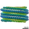 7343MC M: map data used to model this data C: citing same article ( |
|---|---|
| Similar structure data |

-Links
 PDBj
PDBj
- Assembly
| Deposited unit | 
|
|---|---|
| 1 | x 60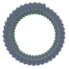
|
| 2 |
|
| 3 | 
|
| Symmetry | Helical symmetry: (Circular symmetry: 1 / Dyad axis: no / N subunits divisor: 1 / Num. of operations: 60 / Rise per n subunits: 2.65 Å / Rotation per n subunits: -8.53 °) |
-Components
| #1: Protein | Mass: 48142.590 Da / Num. of mol.: 2 / Fragment: UNP residues 25-457 Source method: isolated from a genetically manipulated source Source: (gene. exp.) Zaire ebolavirus / Gene: NP / Production host:  |
|---|
-Experimental details
-Experiment
| Experiment | Method: ELECTRON MICROSCOPY |
|---|---|
| EM experiment | Aggregation state: FILAMENT / 3D reconstruction method: helical reconstruction |
- Sample preparation
| Component | Name: eNP nucleocapsid-like assembly / Type: COMPLEX / Entity ID: all / Source: RECOMBINANT |
|---|---|
| Molecular weight | Experimental value: NO |
| Source (natural) | Organism: Zaire ebolavirus |
| Source (recombinant) | Organism: |
| Buffer solution | pH: 7.4 / Details: 150 mM NaCl, 3 mM KCl, 10 mM Na2HPO4, 2 mM KH2PO4 |
| Buffer component | Conc.: 12 mM / Name: Phosphate-buffered saline / Formula: PBS |
| Specimen | Conc.: 5 mg/ml / Embedding applied: NO / Shadowing applied: NO / Staining applied: NO / Vitrification applied: YES |
| Specimen support | Grid material: COPPER / Grid mesh size: 200 divisions/in. / Grid type: Quantifoil R1.2/1.3 |
| Vitrification | Instrument: FEI VITROBOT MARK IV / Cryogen name: ETHANE / Humidity: 100 % / Chamber temperature: 283 K |
- Electron microscopy imaging
| Microscopy | Model: JEOL 3200FSC |
|---|---|
| Electron gun | Electron source:  FIELD EMISSION GUN / Accelerating voltage: 300 kV / Illumination mode: FLOOD BEAM FIELD EMISSION GUN / Accelerating voltage: 300 kV / Illumination mode: FLOOD BEAM |
| Electron lens | Mode: BRIGHT FIELD / Nominal magnification: 30000 X / Cs: 4.7 mm / C2 aperture diameter: 70 µm / Alignment procedure: BASIC |
| Specimen holder | Cryogen: NITROGEN / Specimen holder model: JEOL 3200FSC CRYOHOLDER / Temperature (max): 86 K / Temperature (min): 86 K |
| Image recording | Average exposure time: 8 sec. / Electron dose: 24 e/Å2 / Detector mode: COUNTING / Film or detector model: GATAN K2 SUMMIT (4k x 4k) / Num. of grids imaged: 2 / Num. of real images: 1266 |
| EM imaging optics | Energyfilter name: In-column Omega Filter / Energyfilter upper: 30 eV / Energyfilter lower: 30 eV |
| Image scans | Width: 3838 / Height: 3710 / Movie frames/image: 40 / Used frames/image: 2-32 |
- Processing
| Software | Name: PHENIX / Version: 1.13_2998: / Classification: refinement | |||||||||||||||||||||||||||||||||||
|---|---|---|---|---|---|---|---|---|---|---|---|---|---|---|---|---|---|---|---|---|---|---|---|---|---|---|---|---|---|---|---|---|---|---|---|---|
| EM software |
| |||||||||||||||||||||||||||||||||||
| CTF correction | Type: PHASE FLIPPING AND AMPLITUDE CORRECTION | |||||||||||||||||||||||||||||||||||
| Helical symmerty | Angular rotation/subunit: -8.53 ° / Axial rise/subunit: 2.65 Å / Axial symmetry: C1 | |||||||||||||||||||||||||||||||||||
| Particle selection | Num. of particles selected: 200685 | |||||||||||||||||||||||||||||||||||
| 3D reconstruction | Resolution: 5.8 Å / Resolution method: FSC 0.143 CUT-OFF / Num. of particles: 169526 / Symmetry type: HELICAL | |||||||||||||||||||||||||||||||||||
| Atomic model building | B value: 276 / Protocol: FLEXIBLE FIT / Space: REAL / Target criteria: Correlation coefficient | |||||||||||||||||||||||||||||||||||
| Refine LS restraints |
|
