 Movie
Movie Controller

[English] 日本語
 Yorodumi
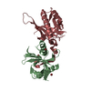
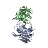
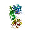
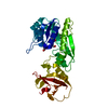
- PDB-6taw: 3C-like protease from Southampton virus complexed with FMOPL000411a. -
+ Open data

- Basic information
| Entry | Database: PDB / ID: 6taw | ||||||
|---|---|---|---|---|---|---|---|
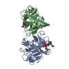
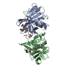
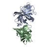
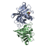
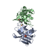
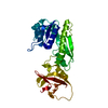
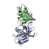
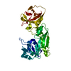
| Title | 3C-like protease from Southampton virus complexed with FMOPL000411a. | ||||||
 Components Components | (Genome polyprotein) x 2 | ||||||
 Keywords Keywords | HYDROLASE / Viral protease. | ||||||
| Function / homology |  Function and homology information Function and homology informationcalicivirin / host cell Golgi membrane / ribonucleoside triphosphate phosphatase activity / nucleoside-triphosphate phosphatase / RNA helicase activity / host cell endoplasmic reticulum membrane / RNA-directed RNA polymerase / cysteine-type endopeptidase activity / viral RNA genome replication / RNA-directed RNA polymerase activity ...calicivirin / host cell Golgi membrane / ribonucleoside triphosphate phosphatase activity / nucleoside-triphosphate phosphatase / RNA helicase activity / host cell endoplasmic reticulum membrane / RNA-directed RNA polymerase / cysteine-type endopeptidase activity / viral RNA genome replication / RNA-directed RNA polymerase activity / DNA-templated transcription / proteolysis / RNA binding / ATP binding / metal ion binding / membrane Similarity search - Function | ||||||
| Biological species | Southampton virus | ||||||
| Method |  X-RAY DIFFRACTION / SYNCHROTRON / FOURIER SYNTHESIS / Resolution: 1.41 Å X-RAY DIFFRACTION / SYNCHROTRON / FOURIER SYNTHESIS / Resolution: 1.41 Å | ||||||
 Authors Authors | Guo, J. / Cooper, J.B. | ||||||
 Citation Citation | Journal: J Struct Biol X / Year: 2020 Title: In crystallo-screening for discovery of human norovirus 3C-like protease inhibitors. Authors: Guo, J. / Douangamath, A. / Song, W. / Coker, A.R. / Chan, A.W.E. / Wood, S.P. / Cooper, J.B. / Resnick, E. / London, N. / Delft, F.V. | ||||||
| History |
|
- Structure visualization
| Structure viewer | Molecule: MolmilJmol/JSmol |
|---|
- Downloads & links
-Download
| PDBx/mmCIF format | 6taw.cif.gz | 168.1 KB | Display | PDBx/mmCIF format |
|---|---|---|---|---|
| PDB format | pdb6taw.ent.gz | 132 KB | Display | PDB format |
| PDBx/mmJSON format | 6taw.json.gz | Tree view | PDBx/mmJSON format | |
| Others |  Other downloads Other downloads |
-Validation report
| Summary document | 6taw_validation.pdf.gz | 681.1 KB | Display | wwPDB validaton report |
|---|---|---|---|---|
| Full document | 6taw_full_validation.pdf.gz | 683.6 KB | Display | |
| Data in XML | 6taw_validation.xml.gz | 19.5 KB | Display | |
| Data in CIF | 6taw_validation.cif.gz | 29.9 KB | Display | |
| Arichive directory | https://data.pdbj.org/pub/pdb/validation_reports/ta/6tawftp://data.pdbj.org/pub/pdb/validation_reports/ta/6taw | HTTPS FTP |
-Related structure data
| Related structure data | 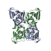 6t1qSC 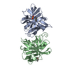 6t2iC  6t2xC  6t3gC  6t49C  6t4eC  6t4sC 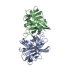 6t5dC 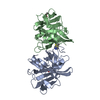 6t5rC  6t6wC  6t71C 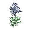 6t82C  6t8rC 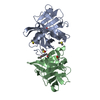 6t8tC 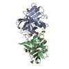 6talC  6tboC  6tbpC 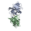 6tc1C 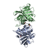 6tcfC  6tglC S: Starting model for refinement C: citing same article ( |
|---|---|
| Similar structure data |

-Links
 PDBj
PDBj
- Assembly
| Deposited unit | 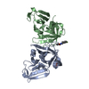
| ||||||||||||
|---|---|---|---|---|---|---|---|---|---|---|---|---|---|
| 1 |
| ||||||||||||
| Unit cell |
| ||||||||||||
| Components on special symmetry positions |
|
-Components
| #1: Protein | Mass: 18387.350 Da / Num. of mol.: 1 Source method: isolated from a genetically manipulated source Source: (gene. exp.)  Southampton virus (serotype 3) / Strain: serotype 3 / Gene: ORF1 Southampton virus (serotype 3) / Strain: serotype 3 / Gene: ORF1Production host:  References: UniProt: Q04544, nucleoside-triphosphate phosphatase, calicivirin, RNA-directed RNA polymerase |
|---|---|
| #2: Protein | Mass: 18290.234 Da / Num. of mol.: 1 Source method: isolated from a genetically manipulated source Source: (gene. exp.) Southampton virus (serotype 3) / Strain: serotype 3 / Gene: ORF1Production host: References: UniProt: Q04544, nucleoside-triphosphate phosphatase, calicivirin, RNA-directed RNA polymerase |
| #3: Chemical | ChemComp-N08 / ~{  Mass: 233.238 Da / Num. of mol.: 1 / Source method: obtained synthetically / Formula: C13H12FNO2 / Feature type: SUBJECT OF INVESTIGATION Mass: 233.238 Da / Num. of mol.: 1 / Source method: obtained synthetically / Formula: C13H12FNO2 / Feature type: SUBJECT OF INVESTIGATION |
| #4: Chemical | ChemComp-PO4 /   Mass: 94.971 Da / Num. of mol.: 1 / Source method: obtained synthetically / Formula: PO4 Mass: 94.971 Da / Num. of mol.: 1 / Source method: obtained synthetically / Formula: PO4 |
| #5: Water | ChemComp-HOH /  Mass: 18.015 Da / Num. of mol.: 423 / Source method: isolated from a natural source / Formula: H2O Mass: 18.015 Da / Num. of mol.: 423 / Source method: isolated from a natural source / Formula: H2O |
| Has ligand of interest | Y |
-Experimental details
-Experiment
| Experiment | Method: X-RAY DIFFRACTION / Number of used crystals: 1 |
|---|
- Sample preparation
| Crystal | Density Matthews: 2.32 Å3/Da / Density % sol: 47.08 % |
|---|---|
| Crystal grow | Temperature: 294 K / Method: vapor diffusion, sitting drop / pH: 5.1 Details: Protein concentration 4 mg/ml. 0.2 M ammonium citrate and 12% (v/v) PEG3350. |
-Data collection
| Diffraction | Mean temperature: 100 K / Serial crystal experiment: N | |||||||||||||||||||||||||||
|---|---|---|---|---|---|---|---|---|---|---|---|---|---|---|---|---|---|---|---|---|---|---|---|---|---|---|---|---|
| Diffraction source | Source: SYNCHROTRON / Site: Diamond  / Beamline: I04-1 / Wavelength: 0.92819 Å / Beamline: I04-1 / Wavelength: 0.92819 Å | |||||||||||||||||||||||||||
| Detector | Type: DECTRIS PILATUS 6M / Detector: PIXEL / Date: Dec 4, 2016 | |||||||||||||||||||||||||||
| Radiation | Protocol: SINGLE WAVELENGTH / Monochromatic (M) / Laue (L): M / Scattering type: x-ray | |||||||||||||||||||||||||||
| Radiation wavelength | Wavelength: 0.92819 Å / Relative weight: 1 | |||||||||||||||||||||||||||
| Reflection | Resolution: 1.41→51.17 Å / Num. obs: 61836 / % possible obs: 96 % / Redundancy: 3 % / CC1/2: 0.999 / Rmerge(I) obs: 0.041 / Rpim(I) all: 0.027 / Rrim(I) all: 0.049 / Net I/σ(I): 10.4 | |||||||||||||||||||||||||||
| Reflection shell | Diffraction-ID: 1
|
- Processing
| Software |
| |||||||||||||||||||||||||||||||||||||||||||||||||||||||||||||||||
|---|---|---|---|---|---|---|---|---|---|---|---|---|---|---|---|---|---|---|---|---|---|---|---|---|---|---|---|---|---|---|---|---|---|---|---|---|---|---|---|---|---|---|---|---|---|---|---|---|---|---|---|---|---|---|---|---|---|---|---|---|---|---|---|---|---|---|
| Refinement | Method to determine structure: FOURIER SYNTHESIS Starting model: 6t1q Resolution: 1.41→51.17 Å / Cor.coef. Fo:Fc: 0.985 / Cor.coef. Fo:Fc free: 0.972 / SU B: 3.219 / SU ML: 0.051 / SU R Cruickshank DPI: 0.0593 / Cross valid method: THROUGHOUT / σ(F): 0 / ESU R: 0.059 / ESU R Free: 0.06 Details: HYDROGENS HAVE BEEN ADDED IN THE RIDING POSITIONS ANISOTROPIC U VALUES : REFINED INDIVIDUALLY
| |||||||||||||||||||||||||||||||||||||||||||||||||||||||||||||||||
| Solvent computation | Ion probe radii: 0.8 Å / Shrinkage radii: 0.8 Å / VDW probe radii: 1.2 Å | |||||||||||||||||||||||||||||||||||||||||||||||||||||||||||||||||
| Displacement parameters | Biso max: 149.47 Å2 / Biso mean: 24.218 Å2 / Biso min: 9.45 Å2
| |||||||||||||||||||||||||||||||||||||||||||||||||||||||||||||||||
| Refinement step | Cycle: final / Resolution: 1.41→51.17 Å
| |||||||||||||||||||||||||||||||||||||||||||||||||||||||||||||||||
| Refine LS restraints |
| |||||||||||||||||||||||||||||||||||||||||||||||||||||||||||||||||
| LS refinement shell | Resolution: 1.41→1.446 Å / Rfactor Rfree error: 0 / Total num. of bins used: 20
|
