 Movie
Movie Controller

[English] 日本語
 Yorodumi
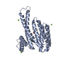
- PDB-6hkb: Ternary complex of Estrogen Receptor alpha peptide and 14-3-3 sig... -
+ Open data

- Basic information
| Entry | Database: PDB / ID: 6hkb | ||||||
|---|---|---|---|---|---|---|---|
| Title | Ternary complex of Estrogen Receptor alpha peptide and 14-3-3 sigma C42 mutant bound to disulfide fragment PPI stabilizer 3 | ||||||
 Components Components |
| ||||||
 Keywords Keywords | PROTEIN BINDING / protein-protein interaction / fragment / tethering / stabilizer | ||||||
| Function / homology |  Function and homology information Function and homology informationsteroid hormone receptor signaling pathway / RUNX1 regulates transcription of genes involved in WNT signaling / RUNX1 regulates estrogen receptor mediated transcription / nuclear estrogen receptor activity / positive regulation of epidermal cell differentiation / regulation of epidermal cell division / protein kinase C inhibitor activity / Developmental Lineage of Mammary Gland Alveolar Cells / positive regulation of hippo signaling / regulation of cell-cell adhesion ...steroid hormone receptor signaling pathway / RUNX1 regulates transcription of genes involved in WNT signaling / RUNX1 regulates estrogen receptor mediated transcription / nuclear estrogen receptor activity / positive regulation of epidermal cell differentiation / regulation of epidermal cell division / protein kinase C inhibitor activity / Developmental Lineage of Mammary Gland Alveolar Cells / positive regulation of hippo signaling / regulation of cell-cell adhesion / TFIIB-class transcription factor binding / negative regulation of smooth muscle cell apoptotic process / establishment of skin barrier / Regulation of localization of FOXO transcription factors / nuclear receptor-mediated steroid hormone signaling pathway / Regulation of GBP-mediated host defense / cellular response to estrogen stimulus / Developmental Lineage of Mammary Gland Luminal Epithelial Cells / estrogen response element binding / positive regulation of keratinocyte differentiation / phosphoserine residue binding / Activation of BAD and translocation to mitochondria / Mitochondrial unfolded protein response (UPRmt) / negative regulation of protein localization to plasma membrane / Nuclear signaling by ERBB4 / cAMP/PKA signal transduction / protein export from nucleus / estrogen receptor signaling pathway / SARS-CoV-2 targets host intracellular signalling and regulatory pathways / RNA polymerase II preinitiation complex assembly / SARS-CoV-1 targets host intracellular signalling and regulatory pathways / release of cytochrome c from mitochondria / RHO GTPases activate PKNs / Chk1/Chk2(Cds1) mediated inactivation of Cyclin B:Cdk1 complex / positive regulation of nitric-oxide synthase activity / steroid binding / negative regulation of protein kinase activity / stem cell differentiation / positive regulation of protein export from nucleus / protein localization to chromatin / TFAP2 (AP-2) family regulates transcription of growth factors and their receptors / keratinocyte differentiation / positive regulation of protein localization / 14-3-3 protein binding / ESR-mediated signaling / negative regulation of miRNA transcription / positive regulation of cell adhesion / TP53 Regulates Transcription of Genes Involved in G2 Cell Cycle Arrest / negative regulation of innate immune response / TBP-class protein binding / nitric-oxide synthase regulator activity / nuclear estrogen receptor binding / transcription corepressor binding / transcription coregulator binding / negative regulation of canonical NF-kappaB signal transduction / cellular response to estradiol stimulus / SUMOylation of intracellular receptors / TP53 Regulates Metabolic Genes / intrinsic apoptotic signaling pathway in response to DNA damage / Translocation of SLC2A4 (GLUT4) to the plasma membrane / euchromatin / Nuclear Receptor transcription pathway / protein sequestering activity / response to estrogen / beta-catenin binding / nuclear receptor activity / intracellular protein localization / positive regulation of nitric oxide biosynthetic process / Constitutive Signaling by Aberrant PI3K in Cancer / sequence-specific double-stranded DNA binding / transcription coactivator binding / phospholipase C-activating G protein-coupled receptor signaling pathway / Regulation of RUNX2 expression and activity / response to estradiol / Ovarian tumor domain proteases / PIP3 activates AKT signaling / regulation of protein localization / positive regulation of cell growth / ATPase binding / PI5P, PP2A and IER3 Regulate PI3K/AKT Signaling / positive regulation of cytosolic calcium ion concentration / DNA-binding transcription activator activity, RNA polymerase II-specific / transcription regulator complex / Estrogen-dependent gene expression / calmodulin binding / DNA-binding transcription factor activity, RNA polymerase II-specific / Extra-nuclear estrogen signaling / chromatin remodeling / RNA polymerase II cis-regulatory region sequence-specific DNA binding / cadherin binding / DNA-binding transcription factor activity / negative regulation of gene expression / chromatin binding / regulation of transcription by RNA polymerase II / regulation of DNA-templated transcription / positive regulation of DNA-templated transcription / protein kinase binding / chromatin / negative regulation of transcription by RNA polymerase II / Golgi apparatus Similarity search - Function | ||||||
| Biological species |  Homo sapiens (human) Homo sapiens (human) | ||||||
| Method |  X-RAY DIFFRACTION / MOLECULAR REPLACEMENT / Resolution: 1.7 Å X-RAY DIFFRACTION / MOLECULAR REPLACEMENT / Resolution: 1.7 Å | ||||||
 Authors Authors | Sijbesma, E. / Hallenbeck, K.K. / Leysen, S. / Arkin, M.R. / Ottmann, C. | ||||||
| Funding support |  Netherlands, 1items Netherlands, 1items
| ||||||
 Citation Citation | Journal: J. Am. Chem. Soc. / Year: 2019 Title: Site-Directed Fragment-Based Screening for the Discovery of Protein-Protein Interaction Stabilizers. Authors: Sijbesma, E. / Hallenbeck, K.K. / Leysen, S. / de Vink, P.J. / Skora, L. / Jahnke, W. / Brunsveld, L. / Arkin, M.R. / Ottmann, C. | ||||||
| History |
|
- Structure visualization
| Structure viewer | Molecule: MolmilJmol/JSmol |
|---|
- Downloads & links
-Download
| PDBx/mmCIF format | 6hkb.cif.gz | 73 KB | Display | PDBx/mmCIF format |
|---|---|---|---|---|
| PDB format | pdb6hkb.ent.gz | 50.7 KB | Display | PDB format |
| PDBx/mmJSON format | 6hkb.json.gz | Tree view | PDBx/mmJSON format | |
| Others |  Other downloads Other downloads |
-Validation report
| Arichive directory | https://data.pdbj.org/pub/pdb/validation_reports/hk/6hkbftp://data.pdbj.org/pub/pdb/validation_reports/hk/6hkb | HTTPS FTP |
|---|
-Related structure data
| Related structure data | 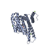 6hhpC 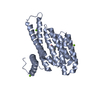 6hkfC 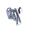 6hmtC 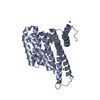 6hmuC  6hn2C 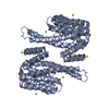 4jc3S S: Starting model for refinement C: citing same article ( |
|---|---|
| Similar structure data |

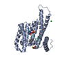
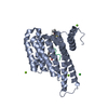

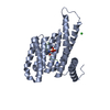
-Links
 PDBj
PDBj



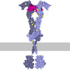











- Assembly
| Deposited unit | 
| ||||||||
|---|---|---|---|---|---|---|---|---|---|
| 1 | 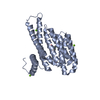
| ||||||||
| Unit cell |
| ||||||||
| Components on special symmetry positions |
|
-Components
| #1: Protein | Mass: 26542.914 Da / Num. of mol.: 1 / Mutation: C38N, N42C Source method: isolated from a genetically manipulated source Source: (gene. exp.) Homo sapiens (human) / Gene: SFN, HME1 / Production host:   Escherichia coli (E. coli) / References: UniProt: P31947 Escherichia coli (E. coli) / References: UniProt: P31947 | ||||||
|---|---|---|---|---|---|---|---|
| #2: Protein/peptide | Mass: 870.840 Da / Num. of mol.: 1 / Source method: obtained synthetically / Source: (synth.) Homo sapiens (human) / References: UniProt: P03372 | ||||||
| #3: Chemical |   Mass: 24.305 Da / Num. of mol.: 3 / Source method: obtained synthetically / Formula: Mg Mass: 24.305 Da / Num. of mol.: 3 / Source method: obtained synthetically / Formula: Mg#4: Chemical | ChemComp-G8Q / ( | 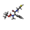 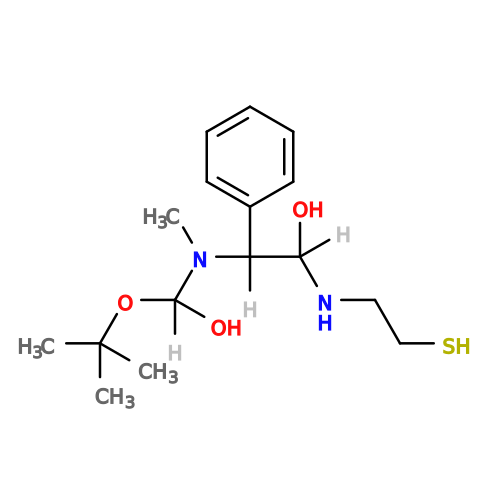 Mass: 328.470 Da / Num. of mol.: 1 / Source method: obtained synthetically / Formula: C16H28N2O3S / Feature type: SUBJECT OF INVESTIGATION Mass: 328.470 Da / Num. of mol.: 1 / Source method: obtained synthetically / Formula: C16H28N2O3S / Feature type: SUBJECT OF INVESTIGATION#5: Water | ChemComp-HOH / |  Mass: 18.015 Da / Num. of mol.: 339 / Source method: isolated from a natural source / Formula: H2O Mass: 18.015 Da / Num. of mol.: 339 / Source method: isolated from a natural source / Formula: H2OHas protein modification | Y | |
-Experimental details
-Experiment
| Experiment | Method: X-RAY DIFFRACTION / Number of used crystals: 1 |
|---|
- Sample preparation
| Crystal | Density Matthews: 2.63 Å3/Da / Density % sol: 53.26 % |
|---|---|
| Crystal grow | Temperature: 278 K / Method: vapor diffusion, sitting drop / pH: 7.1 / Details: 0.095M Hepes, 0.19M CaCl2, 5% glycerol, 26% PEG400 |
-Data collection
| Diffraction | Mean temperature: 100 K |
|---|---|
| Diffraction source | Source: SEALED TUBE / Type: RIGAKU MICROMAX-003 / Wavelength: 1.54 Å |
| Detector | Type: DECTRIS PILATUS 200K / Detector: PIXEL / Date: Mar 11, 2016 |
| Radiation | Protocol: SINGLE WAVELENGTH / Monochromatic (M) / Laue (L): M / Scattering type: x-ray |
| Radiation wavelength | Wavelength: 1.54 Å / Relative weight: 1 |
| Reflection | Resolution: 1.7→66.37 Å / Num. obs: 32176 / % possible obs: 99.9 % / Redundancy: 4.7 % / Biso Wilson estimate: 10.79 Å2 / CC1/2: 0.995 / Rrim(I) all: 0.084 / Rsym value: 0.075 / Net I/σ(I): 15.4 |
| Reflection shell | Resolution: 1.7→1.73 Å / Redundancy: 4.6 % / Mean I/σ(I) obs: 7.5 / Num. unique obs: 1681 / CC1/2: 0.967 / Rrim(I) all: 0.212 / Rsym value: 0.188 / % possible all: 100 |
- Processing
| Software |
| |||||||||||||||||||||||||||||||||||||||||||||||||||||||||||||||||||||||||||||||||||||||||||
|---|---|---|---|---|---|---|---|---|---|---|---|---|---|---|---|---|---|---|---|---|---|---|---|---|---|---|---|---|---|---|---|---|---|---|---|---|---|---|---|---|---|---|---|---|---|---|---|---|---|---|---|---|---|---|---|---|---|---|---|---|---|---|---|---|---|---|---|---|---|---|---|---|---|---|---|---|---|---|---|---|---|---|---|---|---|---|---|---|---|---|---|---|
| Refinement | Method to determine structure: MOLECULAR REPLACEMENT Starting model: 4JC3 Resolution: 1.7→34.34 Å / SU ML: 0.14 / Cross valid method: THROUGHOUT / σ(F): 1.34 / Phase error: 18.45
| |||||||||||||||||||||||||||||||||||||||||||||||||||||||||||||||||||||||||||||||||||||||||||
| Solvent computation | Shrinkage radii: 0.9 Å / VDW probe radii: 1.11 Å | |||||||||||||||||||||||||||||||||||||||||||||||||||||||||||||||||||||||||||||||||||||||||||
| Displacement parameters | Biso max: 58.97 Å2 / Biso mean: 13.7022 Å2 / Biso min: 3.82 Å2 | |||||||||||||||||||||||||||||||||||||||||||||||||||||||||||||||||||||||||||||||||||||||||||
| Refinement step | Cycle: final / Resolution: 1.7→34.34 Å
| |||||||||||||||||||||||||||||||||||||||||||||||||||||||||||||||||||||||||||||||||||||||||||
| Refine LS restraints |
| |||||||||||||||||||||||||||||||||||||||||||||||||||||||||||||||||||||||||||||||||||||||||||
| LS refinement shell | Refine-ID: X-RAY DIFFRACTION / Rfactor Rfree error: 0 / Total num. of bins used: 12
|
