 Movie
Movie Controller

[English] 日本語
 Yorodumi
- EMDB-6828: Cryo-EM structure of the RC-LH core complex from Roseiflexus cast... -
+ Open data

- Basic information
| Entry | Database: EMDB / ID: EMD-6828 | |||||||||
|---|---|---|---|---|---|---|---|---|---|---|
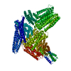
| Title | Cryo-EM structure of the RC-LH core complex from Roseiflexus castenholzii | |||||||||
 Map data Map data | ||||||||||
 Sample Sample |
| |||||||||
| Function / homology |  Function and homology information Function and homology informationorganelle inner membrane / plasma membrane light-harvesting complex / bacteriochlorophyll binding / photosynthetic electron transport in photosystem II / photosynthesis, light reaction / endomembrane system / electron transfer activity / iron ion binding / heme binding / metal ion binding / plasma membrane Similarity search - Function | |||||||||
| Biological species |  Roseiflexus castenholzii (bacteria) Roseiflexus castenholzii (bacteria) | |||||||||
| Method | single particle reconstruction / cryo EM / Resolution: 4.1 Å | |||||||||
 Authors Authors | Xin YY / Shi Y / Niu TX / Wang QQ / Niu WQ / Huang XJ / Ding W / Blankenship RE / Xu XL / Sun F | |||||||||
 Citation Citation | Journal: Nat Commun / Year: 2018 Title: Cryo-EM structure of the RC-LH core complex from an early branching photosynthetic prokaryote. Authors: Yueyong Xin / Yang Shi / Tongxin Niu / Qingqiang Wang / Wanqiang Niu / Xiaojun Huang / Wei Ding / Lei Yang / Robert E Blankenship / Xiaoling Xu / Fei Sun /   Abstract: Photosynthetic prokaryotes evolved diverse light-harvesting (LH) antennas to absorb sunlight and transfer energy to reaction centers (RC). The filamentous anoxygenic phototrophs (FAPs) are important ...Photosynthetic prokaryotes evolved diverse light-harvesting (LH) antennas to absorb sunlight and transfer energy to reaction centers (RC). The filamentous anoxygenic phototrophs (FAPs) are important early branching photosynthetic bacteria in understanding the origin and evolution of photosynthesis. How their photosynthetic machinery assembles for efficient energy transfer is yet to be elucidated. Here, we report the 4.1 Å structure of photosynthetic core complex from Roseiflexus castenholzii by cryo-electron microscopy. The RC-LH complex has a tetra-heme cytochrome c bound RC encompassed by an elliptical LH ring that is assembled from 15 LHαβ subunits. An N-terminal transmembrane helix of cytochrome c inserts into the LH ring, not only yielding a tightly bound cytochrome c for rapid electron transfer, but also opening a slit in the LH ring, which is further flanked by a transmembrane helix from a newly discovered subunit X. These structural features suggest an unusual quinone exchange model of prokaryotic photosynthetic machinery. | |||||||||
| History |
|
- Structure visualization
| Movie |
Movie viewer |
|---|---|
| Structure viewer | EM map: SurfViewMolmilJmol/JSmol |
| Supplemental images |

- Downloads & links
-EMDB archive
| Map data | emd_6828.map.gz | 25.1 MB | EMDB map data format | |
|---|---|---|---|---|
| Header (meta data) | emd-6828-v30.xmlemd-6828.xml | 13.2 KB 13.2 KB | Display Display | EMDB header |
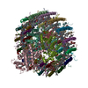
| Images | 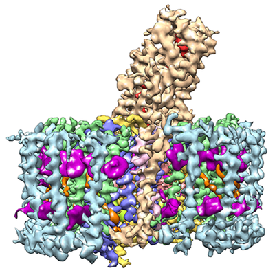 emd_6828.png emd_6828.png | 217.1 KB | ||
| Archive directory |  http://ftp.pdbj.org/pub/emdb/structures/EMD-6828ftp://ftp.pdbj.org/pub/emdb/structures/EMD-6828 http://ftp.pdbj.org/pub/emdb/structures/EMD-6828ftp://ftp.pdbj.org/pub/emdb/structures/EMD-6828 | HTTPS FTP |
-Related structure data
| Related structure data |  5yq7MC M: atomic model generated by this map C: citing same article ( |
|---|---|
| Similar structure data |

-Links
| EMDB pages | EMDB (EBI/PDBe) / EMDataResource |
|---|---|
| Related items in Molecule of the Month |


-Map
| File | Download / File: emd_6828.map.gz / Format: CCP4 / Size: 27 MB / Type: IMAGE STORED AS FLOATING POINT NUMBER (4 BYTES) | ||||||||||||||||||||||||||||||||||||||||||||||||||||||||||||
|---|---|---|---|---|---|---|---|---|---|---|---|---|---|---|---|---|---|---|---|---|---|---|---|---|---|---|---|---|---|---|---|---|---|---|---|---|---|---|---|---|---|---|---|---|---|---|---|---|---|---|---|---|---|---|---|---|---|---|---|---|---|
| Projections & slices | Image control
Images are generated by Spider. | ||||||||||||||||||||||||||||||||||||||||||||||||||||||||||||
| Voxel size | X=Y=Z: 1.12 Å | ||||||||||||||||||||||||||||||||||||||||||||||||||||||||||||
| Density |
| ||||||||||||||||||||||||||||||||||||||||||||||||||||||||||||
| Symmetry | Space group: 1 | ||||||||||||||||||||||||||||||||||||||||||||||||||||||||||||
| Details | EMDB XML:
CCP4 map header:
| ||||||||||||||||||||||||||||||||||||||||||||||||||||||||||||
 Z (Sec.)
 Y (Row.)
 X (Col.)


-Supplemental data
- Sample components
-Entire : photosynthetic core complex
| Entire | Name: photosynthetic core complex |
|---|---|
| Components |
|
-Supramolecule #1: photosynthetic core complex
| Supramolecule | Name: photosynthetic core complex / type: complex / ID: 1 / Parent: 0 / Macromolecule list: all |
|---|---|
| Source (natural) | Organism: Roseiflexus castenholzii (bacteria) |
| Molecular weight | Theoretical: 330 KDa |
-Macromolecule #1: alpha
| Macromolecule | Name: alpha / type: protein_or_peptide / ID: 1 / Enantiomer: LEVO |
|---|---|
| Source (natural) | Organism: Roseiflexus castenholzii (bacteria) |
| Sequence | String: MKDRPFEFRT SVVVSTLLGL VMALLIHFVV LSSGAFNWLR AP |
-Macromolecule #2: beta
| Macromolecule | Name: beta / type: protein_or_peptide / ID: 2 / Enantiomer: LEVO |
|---|---|
| Source (natural) | Organism: Roseiflexus castenholzii (bacteria) |
| Sequence | String: MTDKPQNDLV PDQWKPLFNN AQWLVHDIVV KTIYGGLIIA VIAHVLCWAW TPWIR |
-Macromolecule #3: LM
| Macromolecule | Name: LM / type: protein_or_peptide / ID: 3 / Enantiomer: LEVO |
|---|---|
| Source (natural) | Organism: Roseiflexus castenholzii (bacteria) |
| Sequence | String: MSAVPRALPL PSGETLPAEA ISSTGSQAAS AEVIPFSIIE EFYKRPGKTL AARFFGVDPF DFWIGRFYVG LFGAISIIGI ILGVAFYLYE GVVNEGTLNI LAMRIEPPPV SQGLNVDPAQ PGFFWFLTMV AATIAFVGWL LRQIDISLKL DMGMEVPIAF GAVVSSWITL ...String: MSAVPRALPL PSGETLPAEA ISSTGSQAAS AEVIPFSIIE EFYKRPGKTL AARFFGVDPF DFWIGRFYVG LFGAISIIGI ILGVAFYLYE GVVNEGTLNI LAMRIEPPPV SQGLNVDPAQ PGFFWFLTMV AATIAFVGWL LRQIDISLKL DMGMEVPIAF GAVVSSWITL QWLRPIAMGA WGHGFPLGIT HHLDWVSNIG YQYYNFFYNP FHAIGITLLF ASTLFLHMHG SAVLSEAKRN ISDQNIHVFW RNILGYSIGE IGIHRVAFWT GAASVLFSNL CIFLSGTFVK DWNAFWGFWD KMPIWNGVGQ GALVAGLSLL GVGLVLGRGR ETPGPIDLHD EEYRDGLEGT IAKPPGHVGW MQRLLGEGQV GPIYVGLWGV ISFITFFASA FIILVDYGRQ VGWNPIIYLR EFWNLAVYPP PTEYGLSWNV PWDKGGAWLA ATFFLHISVL TWWARLYTRA KATGVGTQLA WGFASALSLY FVIYLFHPLA LGNWSAAPGH GFRAILDWTN YVSIHWGNFY YNPFHMLSIF FLLGSTLLLA MHGATIVATS KWKSEMEFTE MMAEGPGTQR AQLFWRWVMG WNANSYNIHI WAWWFAAFTA ITGAIGLFLS GTLVPDWYAW GETAKIVAPW PNPDWAQYVF R |
-Macromolecule #4: cytochrome c
| Macromolecule | Name: cytochrome c / type: protein_or_peptide / ID: 4 / Enantiomer: LEVO |
|---|---|
| Source (natural) | Organism: Roseiflexus castenholzii (bacteria) |
| Sequence | String: MIQQPPTLFP EITNTVRGRF YIVAGIISVV MAVASIAIFW WIFYTITPAP APPLQNPIYV NYTQEPTDYI SAESLAAMNA YIQANPQPQA VQVLKGMTTA QISAYMVAQV SGGLKVDCSY CHNIANFAQQ DGYPNAAKKV TARKMMLMSA DLNQNYTAKL PASVGGYQIT ...String: MIQQPPTLFP EITNTVRGRF YIVAGIISVV MAVASIAIFW WIFYTITPAP APPLQNPIYV NYTQEPTDYI SAESLAAMNA YIQANPQPQA VQVLKGMTTA QISAYMVAQV SGGLKVDCSY CHNIANFAQQ DGYPNAAKKV TARKMMLMSA DLNQNYTAKL PASVGGYQIT CATCHNGKAA GLEPYPIEIM NTLPNDWRLP LELDYPGGLV VTGRKDVSNH EVEQNQFAMY HMNVSMGQGC TFCHNARYFP SYEIAQKNHS IIMLQMTKHI QETYVAPGGR IADGIMAGKS PSCWLCHQGA NIPPGAAKPG QVPAVLSSTP |
-Experimental details
-Structure determination
| Method | cryo EM |
|---|---|
 Processing Processing | single particle reconstruction |
| Aggregation state | particle |
-Sample preparation
| Buffer | pH: 8.5 |
|---|---|
| Grid | Model: Quantifoil R1.2/1.3 / Material: GOLD / Mesh: 300 / Pretreatment - Type: GLOW DISCHARGE / Pretreatment - Atmosphere: AIR |
| Vitrification | Cryogen name: ETHANE / Chamber humidity: 100 % / Chamber temperature: 289 K / Instrument: FEI VITROBOT MARK IV |
- Electron microscopy
| Microscope | FEI TITAN KRIOS |
|---|---|
| Image recording | Film or detector model: FEI FALCON III (4k x 4k) / Detector mode: COUNTING / Average electron dose: 40.0 e/Å2 |
| Electron beam | Acceleration voltage: 300 kV / Electron source:  FIELD EMISSION GUN FIELD EMISSION GUN |
| Electron optics | C2 aperture diameter: 100.0 µm / Calibrated defocus max: 3.5 µm / Calibrated defocus min: 1.0 µm / Calibrated magnification: 73661 / Illumination mode: FLOOD BEAM / Imaging mode: BRIGHT FIELD / Cs: 2.7 mm / Nominal defocus max: 3.5 µm / Nominal defocus min: 1.0 µm / Nominal magnification: 75000 |
| Sample stage | Specimen holder model: FEI TITAN KRIOS AUTOGRID HOLDER |
| Experimental equipment |  Model: Titan Krios / Image courtesy: FEI Company |
-Image processing
| Startup model | Type of model: INSILICO MODEL / In silico model: The startup model was generated by EMAN2. |
|---|---|
| Final reconstruction | Applied symmetry - Point group: C1 (asymmetric) / Resolution.type: BY AUTHOR / Resolution: 4.1 Å / Resolution method: FSC 0.143 CUT-OFF / Number images used: 148618 |
| Initial angle assignment | Type: PROJECTION MATCHING Projection matching processing - Angular sampling: 7.5 degrees |
| Final angle assignment | Type: PROJECTION MATCHING |
