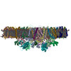
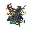
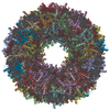
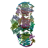
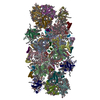
- EMDB-6617: CryoEM map of spinach PSII-LHCII supercomplex at 3.2A resolution -
+
データを開く
IDまたはキーワード:
読み込み中...
-
基本情報
登録情報
データベース: EMDB / ID: EMD-6617
タイトル
CryoEM map of spinach PSII-LHCII supercomplex at 3.2A resolution
マップデータ
A cryo-EM map of a membrane protein complex
試料
試料: Spinach PSII-LHCII supercomplex
タンパク質・ペプチド: Photosystem II (or water-plastoquinone oxidoreductase) in complex with Light harvesting complex II
キーワード
membrane protein / spinach photosystem II / cryo-EM
機能・相同性
機能・相同性情報
chloroplast photosystem II / photosynthesis, light harvesting / plastoglobule / : / photosystem II assembly / photosynthesis, light harvesting in photosystem I / photosystem II oxygen evolving complex / oxygen evolving activity / photosystem II stabilization / photosystem II ...chloroplast photosystem II / photosynthesis, light harvesting / plastoglobule / : / photosystem II assembly / photosynthesis, light harvesting in photosystem I / photosystem II oxygen evolving complex / oxygen evolving activity / photosystem II stabilization / photosystem II / photosystem II reaction center / : / ion binding / chloroplast envelope / oxidoreductase activity, acting on diphenols and related substances as donors, oxygen as acceptor / photosystem I / photosynthetic electron transport chain / response to herbicide / photosystem II / extrinsic component of membrane / chlorophyll binding / photosynthesis, light reaction / electron transporter, transferring electrons within the cyclic electron transport pathway of photosynthesis activity / phosphate ion binding / chloroplast thylakoid membrane / response to light stimulus / photosynthetic electron transport in photosystem II / photosynthesis / phosphoprotein binding / membrane => GO:0016020 / electron transfer activity / protein stabilization / iron ion binding / calcium ion binding / heme binding / metal ion binding 類似検索 - 分子機能
Photosystem II 5kDa protein, chloroplastic / : / PsbP, C-terminal / PsbP / : / Photosystem II PsbW, class 2 / Photosystem II reaction centre W protein (PsbW) / Oxygen-evolving enhancer protein 3 / Oxygen evolving enhancer protein 3 / Mog1/PsbP, alpha/beta/alpha sandwich ...Photosystem II 5kDa protein, chloroplastic / : / PsbP, C-terminal / PsbP / : / Photosystem II PsbW, class 2 / Photosystem II reaction centre W protein (PsbW) / Oxygen-evolving enhancer protein 3 / Oxygen evolving enhancer protein 3 / Mog1/PsbP, alpha/beta/alpha sandwich / PsbQ-like domain superfamily / Photosystem II PsbJ / Photosystem II PsbJ superfamily / PsbJ / Photosystem II PsbO, manganese-stabilising / Manganese-stabilising protein / photosystem II polypeptide / Photosystem II reaction centre M protein (PsbM) / Photosystem II PsbM superfamily / Photosystem II PsbM / Photosystem II PsbZ, reaction centre / Photosystem II PsbZ superfamily / YCF9 / Photosystem II PsbX / Photosystem II reaction centre X protein (PsbX) / Photosystem II PsbT / Photosystem II PsbL / Photosystem II PsbL superfamily / Photosystem II PsbT superfamily / Photosystem II reaction centre T protein / PsbL protein / Photosystem II CP43 reaction centre protein / Photosystem II PsbK / Photosystem II PsbK superfamily / Photosystem II 4 kDa reaction centre component / Photosystem II CP47 reaction centre protein / Photosystem II PsbI / Photosystem II PsbI superfamily / Photosystem II reaction centre I protein (PSII 4.8 kDa protein) / Chlorophyll A-B binding protein, plant and chromista / Photosystem II reaction centre protein H / Photosystem II protein D1 / Photosystem II D2 protein / Photosystem II cytochrome b559, conserved site / Photosystem II cytochrome b559, alpha subunit / Photosystem II cytochrome b559, beta subunit / Photosystem II cytochrome b559, N-terminal / Photosystem II cytochrome b559, alpha subunit, lumenal region / Photosystem II reaction centre protein H superfamily / Photosystem II cytochrome b559, alpha subunit superfamily / Cytochrome b559, alpha (gene psbE) and beta (gene psbF)subunits / Lumenal portion of Cytochrome b559, alpha (gene psbE) subunit / Photosystem II 10 kDa phosphoprotein / Cytochrome b559 subunits heme-binding site signature. / Chlorophyll A-B binding protein / Chlorophyll A-B binding protein / Photosystem antenna protein-like / Photosystem antenna protein-like superfamily / Photosystem II protein / Outer membrane protein/outer membrane enzyme PagP, beta-barrel / Photosynthetic reaction centre, L/M / Photosystem II protein D1/D2 superfamily / Photosynthetic reaction centre protein / Photosynthetic reaction center proteins signature. 類似検索 - ドメイン・相同性
Chlorophyll a-b binding protein, chloroplastic / Chlorophyll a-b binding protein, chloroplastic / Photosystem II 5 kDa protein, chloroplastic / Uncharacterized protein / Chlorophyll a-b binding protein, chloroplastic / Photosystem II CP47 reaction center protein / Photosystem II reaction center protein H / Photosystem II CP43 reaction center protein / Photosystem II D2 protein / Photosystem II reaction center protein K ...Chlorophyll a-b binding protein, chloroplastic / Chlorophyll a-b binding protein, chloroplastic / Photosystem II 5 kDa protein, chloroplastic / Uncharacterized protein / Chlorophyll a-b binding protein, chloroplastic / Photosystem II CP47 reaction center protein / Photosystem II reaction center protein H / Photosystem II CP43 reaction center protein / Photosystem II D2 protein / Photosystem II reaction center protein K / Oxygen-evolving enhancer protein 3, chloroplastic / Oxygen-evolving enhancer protein 2, chloroplastic / Chlorophyll a-b binding protein, chloroplastic / Oxygen-evolving enhancer protein 1, chloroplastic / Cytochrome b559 subunit beta / Photosystem II reaction center protein L / Photosystem II reaction center protein T / Photosystem II reaction center protein I / Photosystem II reaction center protein M / Cytochrome b559 subunit alpha / Photosystem II protein D1 / Photosystem II reaction center W protein, chloroplastic / Photosystem II reaction center protein J / Photosystem II reaction center protein Z 類似検索 - 構成要素
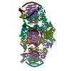
ジャーナル: Nature / 年: 2016 タイトル: Structure of spinach photosystem II-LHCII supercomplex at 3.2 Å resolution. 著者: Xuepeng Wei / Xiaodong Su / Peng Cao / Xiuying Liu / Wenrui Chang / Mei Li / Xinzheng Zhang / Zhenfeng Liu / 要旨: During photosynthesis, the plant photosystem II core complex receives excitation energy from the peripheral light-harvesting complex II (LHCII). The pathways along which excitation energy is ...During photosynthesis, the plant photosystem II core complex receives excitation energy from the peripheral light-harvesting complex II (LHCII). The pathways along which excitation energy is transferred between them, and their assembly mechanisms, remain to be deciphered through high-resolution structural studies. Here we report the structure of a 1.1-megadalton spinach photosystem II-LHCII supercomplex solved at 3.2 Å resolution through single-particle cryo-electron microscopy. The structure reveals a homodimeric supramolecular system in which each monomer contains 25 protein subunits, 105 chlorophylls, 28 carotenoids and other cofactors. Three extrinsic subunits (PsbO, PsbP and PsbQ), which are essential for optimal oxygen-evolving activity of photosystem II, form a triangular crown that shields the Mn4CaO5-binding domains of CP43 and D1. One major trimeric and two minor monomeric LHCIIs associate with each core-complex monomer, and the antenna-core interactions are reinforced by three small intrinsic subunits (PsbW, PsbH and PsbZ). By analysing the closely connected interfacial chlorophylls, we have obtained detailed insights into the energy-transfer pathways between the antenna and core complexes.
分子 #1: Photosystem II (or water-plastoquinone oxidoreductase) in complex...
分子
名称: Photosystem II (or water-plastoquinone oxidoreductase) in complex with Light harvesting complex II タイプ: protein_or_peptide / ID: 1 / 組換発現: No / データベース: NCBI
由来(天然)
生物種: Spinacia oleracea (ホウレンソウ)
-
実験情報
-
構造解析
手法
クライオ電子顕微鏡法
解析
単粒子再構成法
試料の集合状態
particle
-
試料調製
濃度
3 mg/mL
グリッド
詳細: 400 mesh copper grid
凍結
凍結剤: ETHANE / チャンバー内湿度: 100 % / チャンバー内温度: 100 K / 装置: FEI VITROBOT MARK IV
 ムービー
ムービー コントローラー


 データを開く
 基本情報
基本情報 マップデータ
 試料
試料 キーワード
キーワード 機能・相同性情報
 Spinacia oleracea (ホウレンソウ)
 データ登録者
 引用
引用
 構造の表示
構造の表示
 ダウンロードとリンク
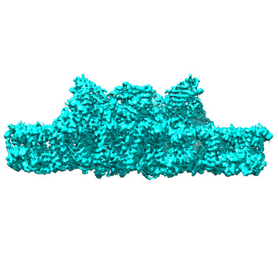 400_6617.gif
 80_6617.gif
 http://ftp.pdbj.org/pub/emdb/structures/EMD-6617
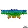


 試料の構成要素
 解析
解析 電子顕微鏡法
 FIELD EMISSION GUN