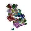
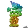
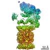
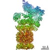
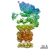
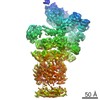
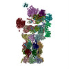
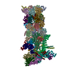
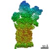
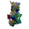
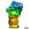
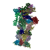
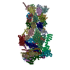
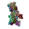
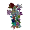
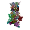
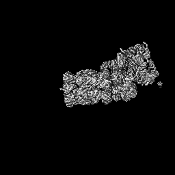
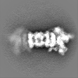
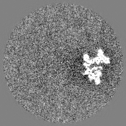
[English] 日本語
Yorodumi- EMDB-6575: Cryo-EM map of yeast 26S proteasome in M2 state derived from Tita... -
+Open data
-Basic information
Entry | Database: EMDB / ID: EMD-6575 | |||||||||
---|---|---|---|---|---|---|---|---|---|---|
Title | Cryo-EM map of yeast 26S proteasome in M2 state derived from Titan dataset | |||||||||
Map data | Reconstruction of single particles | |||||||||
Sample |
| |||||||||
Function / homology | Function and homology information SAGA complex localization to transcription regulatory region / Metalloprotease DUBs / peroxisome fission / proteasome storage granule assembly / transcription export complex 2 / proteasome regulatory particle assembly / protein deneddylation / maintenance of DNA trinucleotide repeats / filamentous growth / COP9 signalosome ...SAGA complex localization to transcription regulatory region / Metalloprotease DUBs / peroxisome fission / proteasome storage granule assembly / transcription export complex 2 / proteasome regulatory particle assembly / protein deneddylation / maintenance of DNA trinucleotide repeats / filamentous growth / COP9 signalosome / proteasome regulatory particle / cytosolic proteasome complex / proteasome regulatory particle, lid subcomplex / protein-containing complex localization / proteasome-activating activity / mitochondrial fission / proteasome regulatory particle, base subcomplex / metal-dependent deubiquitinase activity / nonfunctional rRNA decay / K48-linked polyubiquitin modification-dependent protein binding / proteasome core complex assembly / nuclear outer membrane-endoplasmic reticulum membrane network / Cross-presentation of soluble exogenous antigens (endosomes) / TNFR2 non-canonical NF-kB pathway / proteasomal ubiquitin-independent protein catabolic process / Ubiquitin Mediated Degradation of Phosphorylated Cdc25A / Regulation of PTEN stability and activity / peptide catabolic process / proteasome binding / KEAP1-NFE2L2 pathway / CDK-mediated phosphorylation and removal of Cdc6 / Neddylation / FBXL7 down-regulates AURKA during mitotic entry and in early mitosis / regulation of protein catabolic process / Orc1 removal from chromatin / MAPK6/MAPK4 signaling / proteasome storage granule / Antigen processing: Ubiquitination & Proteasome degradation / polyubiquitin modification-dependent protein binding / endopeptidase activator activity / proteasome assembly / positive regulation of RNA polymerase II transcription preinitiation complex assembly / proteasome endopeptidase complex / proteasome core complex, beta-subunit complex / Ub-specific processing proteases / proteasome core complex, alpha-subunit complex / threonine-type endopeptidase activity / mRNA export from nucleus / enzyme regulator activity / ERAD pathway / Neutrophil degranulation / protein folding chaperone / proteasome complex / ubiquitin binding / nucleotide-excision repair / positive regulation of transcription elongation by RNA polymerase II / double-strand break repair via homologous recombination / positive regulation of protein catabolic process / metallopeptidase activity / peroxisome / protein-macromolecule adaptor activity / ubiquitin-dependent protein catabolic process / proteasome-mediated ubiquitin-dependent protein catabolic process / endopeptidase activity / ubiquitinyl hydrolase 1 / cysteine-type deubiquitinase activity / molecular adaptor activity / regulation of cell cycle / chromatin remodeling / protein domain specific binding / mRNA binding / ubiquitin protein ligase binding / endoplasmic reticulum membrane / structural molecule activity / endoplasmic reticulum / positive regulation of transcription by RNA polymerase II / ATP hydrolysis activity / mitochondrion / ATP binding / identical protein binding / nucleus / metal ion binding / cytosol / cytoplasm Similarity search - Function | |||||||||
Biological species | Saccharomyces cerevisiae (brewer's yeast) | |||||||||
Method | single particle reconstruction / cryo EM / Resolution: 4.6 Å | |||||||||
Authors | Luan B / Huang XL / Wu JP / Shi YG / Wang F | |||||||||
Citation | Journal: Proc Natl Acad Sci U S A / Year: 2016 Title: Structure of an endogenous yeast 26S proteasome reveals two major conformational states. Authors: Bai Luan / Xiuliang Huang / Jianping Wu / Ziqing Mei / Yiwei Wang / Xiaobin Xue / Chuangye Yan / Jiawei Wang / Daniel J Finley / Yigong Shi / Feng Wang / Abstract: The eukaryotic proteasome mediates degradation of polyubiquitinated proteins. Here we report the single-particle cryoelectron microscopy (cryo-EM) structures of the endogenous 26S proteasome from ...The eukaryotic proteasome mediates degradation of polyubiquitinated proteins. Here we report the single-particle cryoelectron microscopy (cryo-EM) structures of the endogenous 26S proteasome from Saccharomyces cerevisiae at 4.6- to 6.3-Å resolution. The fine features of the cryo-EM maps allow modeling of 18 subunits in the regulatory particle and 28 in the core particle. The proteasome exhibits two distinct conformational states, designated M1 and M2, which correspond to those reported previously for the proteasome purified in the presence of ATP-γS and ATP, respectively. These conformations also correspond to those of the proteasome in the presence and absence of exogenous substrate. Structure-guided biochemical analysis reveals enhanced deubiquitylating enzyme activity of Rpn11 upon assembly of the lid. Our structures serve as a molecular basis for mechanistic understanding of proteasome function. | |||||||||
History |
|
-Structure visualization
Movie |
Movie viewer |
---|---|
Structure viewer | EM map: SurfViewMolmilJmol/JSmol |
Supplemental images |
-Downloads & links
-EMDB archive
Map data | emd_6575.map.gz | 59 MB | EMDB map data format | |
---|---|---|---|---|
Header (meta data) | emd-6575-v30.xml emd-6575.xml | 9.7 KB 9.7 KB | Display Display | EMDB header |
Images | 400_6575.gif 80_6575.gif | 55.7 KB 3.8 KB | ||
Archive directory | http://ftp.pdbj.org/pub/emdb/structures/EMD-6575
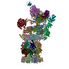
 ftp://ftp.pdbj.org/pub/emdb/structures/EMD-6575 | HTTPS FTP |
-Validation report
Summary document | emd_6575_validation.pdf.gz | 398.4 KB | Display | EMDB validaton report |
---|---|---|---|---|
Full document | emd_6575_full_validation.pdf.gz | 398 KB | Display | |
Data in XML | emd_6575_validation.xml.gz | 6 KB | Display | |
Arichive directory | https://ftp.pdbj.org/pub/emdb/validation_reports/EMD-6575 ftp://ftp.pdbj.org/pub/emdb/validation_reports/EMD-6575 | HTTPS FTP |
-Related structure data
Related structure data | 3jcpMC 6574C 6576C 6577C 6578C 6579C 3jcoC M: atomic model generated by this map C: citing same article (ref.) |
---|---|
Similar structure data |
-Links
EMDB pages | EMDB (EBI/PDBe) / EMDataResource |
---|---|
Related items in Molecule of the Month |
-Map
File | Download / File: emd_6575.map.gz / Format: CCP4 / Size: 62.5 MB / Type: IMAGE STORED AS FLOATING POINT NUMBER (4 BYTES) | ||||||||||||||||||||||||||||||||||||||||||||||||||||||||||||
---|---|---|---|---|---|---|---|---|---|---|---|---|---|---|---|---|---|---|---|---|---|---|---|---|---|---|---|---|---|---|---|---|---|---|---|---|---|---|---|---|---|---|---|---|---|---|---|---|---|---|---|---|---|---|---|---|---|---|---|---|---|
Annotation | Reconstruction of single particles | ||||||||||||||||||||||||||||||||||||||||||||||||||||||||||||
Projections & slices | Image control
Images are generated by Spider. | ||||||||||||||||||||||||||||||||||||||||||||||||||||||||||||
Voxel size | X=Y=Z: 2.1 Å | ||||||||||||||||||||||||||||||||||||||||||||||||||||||||||||
Density |
| ||||||||||||||||||||||||||||||||||||||||||||||||||||||||||||
Symmetry | Space group: 1 | ||||||||||||||||||||||||||||||||||||||||||||||||||||||||||||
Details | EMDB XML:
CCP4 map header:
|
-Supplemental data
-Sample components
-Entire : yeast 26S proteasome in M2 state
Entire | Name: yeast 26S proteasome in M2 state |
---|---|
Components |
|
-Supramolecule #1000: yeast 26S proteasome in M2 state
Supramolecule | Name: yeast 26S proteasome in M2 state / type: sample / ID: 1000 / Details: The sample was monodisperse / Number unique components: 1 |
---|---|
Molecular weight | Experimental: 2.5 MDa / Theoretical: 2.5 MDa |
-Macromolecule #1: 26S proteasome
Macromolecule | Name: 26S proteasome / type: protein_or_peptide / ID: 1 / Number of copies: 1 / Recombinant expression: No / Database: NCBI |
---|---|
Source (natural) | Organism: Saccharomyces cerevisiae (brewer's yeast) / synonym: Yeast / Location in cell: Cytoplasma |
Molecular weight | Experimental: 2.5 MDa / Theoretical: 2.5 MDa |
-Experimental details
-Structure determination
Method | cryo EM |
---|---|
Processing | single particle reconstruction |
Aggregation state | particle |
-Sample preparation
Concentration | 15 mg/mL |
---|---|
Buffer | pH: 7.5 / Details: 50mM Tris7.5, 100mM NaCl, 5mM MgCl2, 2mM ATP |
Grid | Details: Quantifoil Cu R2.0/2.0 200 mesh |
Vitrification | Cryogen name: ETHANE / Chamber humidity: 100 % / Instrument: FEI VITROBOT MARK IV |
-Electron microscopy
Microscope | FEI TITAN KRIOS |
---|---|
Temperature | Average: 100 K |
Date | Nov 2, 2015 |
Image recording | Category: CCD / Film or detector model: FEI FALCON II (4k x 4k) / Number real images: 5517 Details: Every image is the average of 21 frames recorded by the direct electron detector |
Electron beam | Acceleration voltage: 300 kV / Electron source: FIELD EMISSION GUN |
Electron optics | Illumination mode: SPOT SCAN / Imaging mode: BRIGHT FIELD / Cs: 2.7 mm / Nominal defocus max: 0.0025 µm / Nominal defocus min: 0.0015 µm / Nominal magnification: 75000 |
Sample stage | Specimen holder model: FEI TITAN KRIOS AUTOGRID HOLDER |
Experimental equipment | Model: Titan Krios / Image courtesy: FEI Company |
-Image processing
Details | The particles were selected in RELION and manually checked. 3D classification and refinement were performed in RELION. |
---|---|
CTF correction | Details: Each micrographs |
Final reconstruction | Resolution.type: BY AUTHOR / Resolution: 4.6 Å / Resolution method: OTHER / Software - Name: RELION / Number images used: 25151 |
-Atomic model buiding 1
Initial model | PDB ID: |
---|---|
Software | Name: Chimera |
Details | The domains were separately fitted in chimera and then manually checked in Coot. The final model is refined with Phenix. |
Refinement | Space: REAL / Protocol: RIGID BODY FIT |
Output model | PDB-3jcp: |