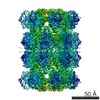
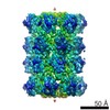
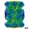
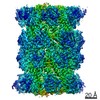
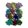
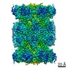
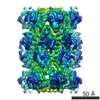
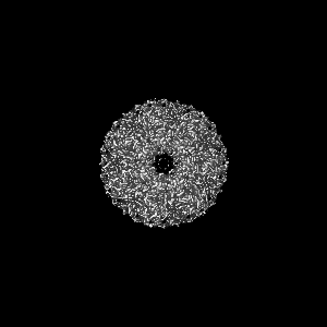
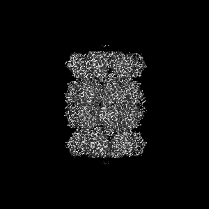
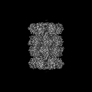
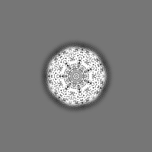
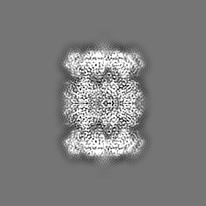
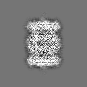
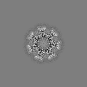
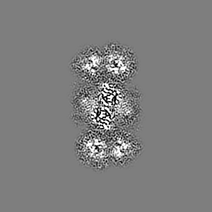
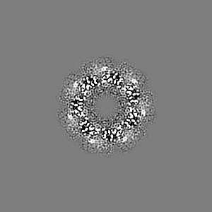
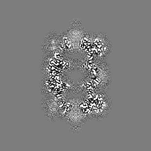
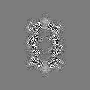
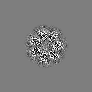
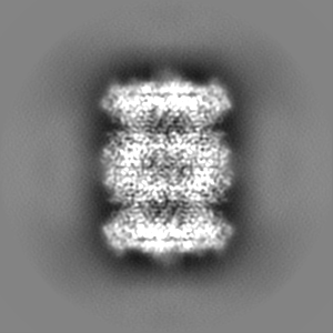
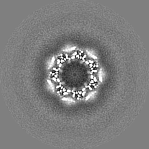
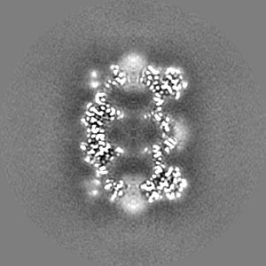
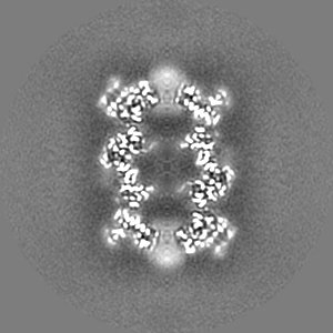
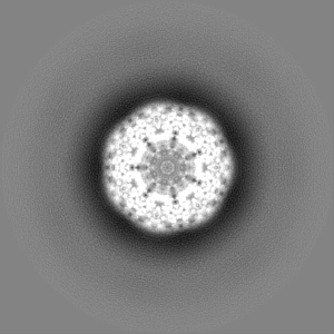
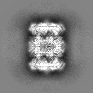
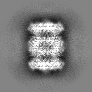
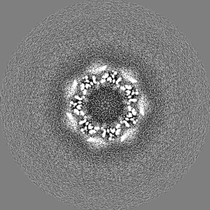
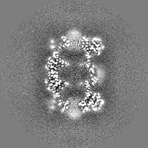
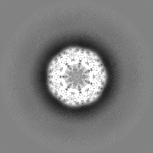
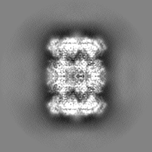
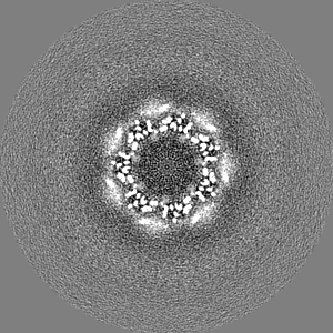
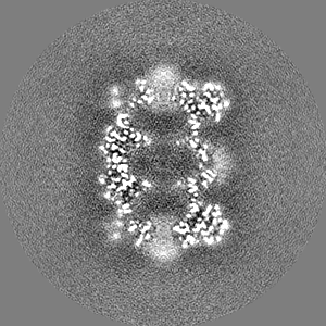
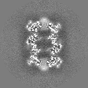
ジャーナル: Elife / 年: 2015 タイトル: 2.8 Å resolution reconstruction of the Thermoplasma acidophilum 20S proteasome using cryo-electron microscopy. 著者: Melody G Campbell / David Veesler / Anchi Cheng / Clinton S Potter / Bridget Carragher / 要旨: Recent developments in detector hardware and image-processing software have revolutionized single particle cryo-electron microscopy (cryoEM) and led to a wave of near-atomic resolution (typically ...Recent developments in detector hardware and image-processing software have revolutionized single particle cryo-electron microscopy (cryoEM) and led to a wave of near-atomic resolution (typically ∼3.3 Å) reconstructions. Reaching resolutions higher than 3 Å is a prerequisite for structure-based drug design and for cryoEM to become widely interesting to pharmaceutical industries. We report here the structure of the 700 kDa Thermoplasma acidophilum 20S proteasome (T20S), determined at 2.8 Å resolution by single-particle cryoEM. The quality of the reconstruction enables identifying the rotameric conformation adopted by some amino-acid side chains (rotamers) and resolving ordered water molecules, in agreement with the expectations for crystal structures at similar resolutions. The results described in this manuscript demonstrate that single particle cryoEM is capable of competing with X-ray crystallography for determination of protein structures of suitable quality for rational drug design.
EMPIAR-10025 (タイトル: T20S Proteasome at 2.8 Å Resolution / Data size: 2.0 TB / Data #1: Raw movies [micrographs - multiframe] Data #2: Frame-averaged micrographs [micrographs - single frame] Data #3: Aligned multi-frame micrographs [micrographs - multiframe])
 ムービー
ムービー コントローラー


 データを開く
 基本情報
基本情報 マップデータ
 試料
試料 キーワード
キーワード 機能・相同性情報
 Thermoplasma acidophilum (好酸性)
 データ登録者
 引用
引用
 構造の表示
構造の表示
 ダウンロードとリンク
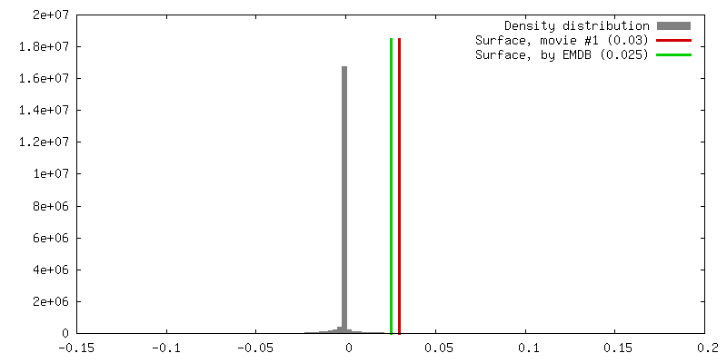
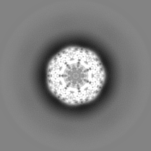
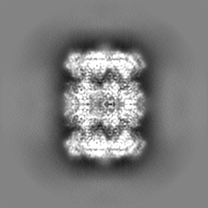
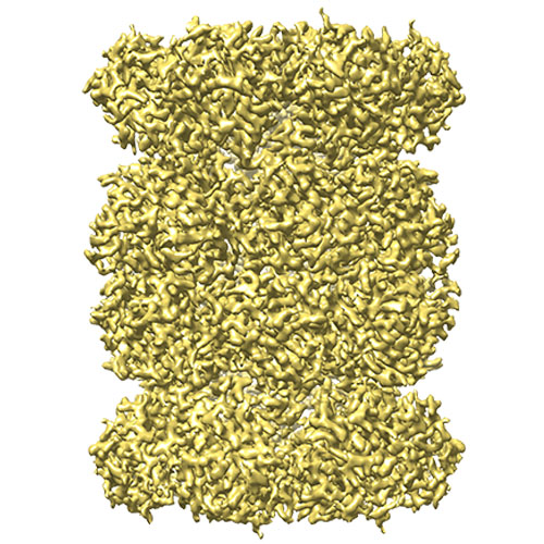 emd_6287.jpg
 http://ftp.pdbj.org/pub/emdb/structures/EMD-6287


 Z (Sec.)
 Y (Row.)
 X (Col.)


 試料の構成要素
 解析
解析 電子顕微鏡法
 FIELD EMISSION GUN
