Movie
Movie Controller

[English] 日本語
 Yorodumi
- PDB-5sv9: Structure of the SLC4 transporter Bor1p in an inward-facing confo... -
+ Open data

- Basic information
| Entry | Database: PDB / ID: 5sv9 | |||||||||||||||
|---|---|---|---|---|---|---|---|---|---|---|---|---|---|---|---|---|
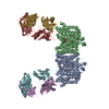
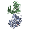
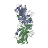
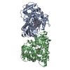
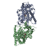
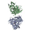
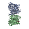
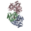
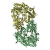
| Title | Structure of the SLC4 transporter Bor1p in an inward-facing conformation | |||||||||||||||
 Components Components | Bor1p boron transporter | |||||||||||||||
 Keywords Keywords | TRANSPORT PROTEIN / boron transporter / anion exchanger family / alternating access mechanism / Structural Genomics / PSI-Biology / Transcontinental EM Initiative for Membrane Protein Structure / TEMIMPS | |||||||||||||||
| Function / homology | Bicarbonate transporter, eukaryotic / Bicarbonate transporter-like, transmembrane domain / HCO3- transporter integral membrane domain / solute:inorganic anion antiporter activity / membrane => GO:0016020 / Bor1p boron transporter Function and homology information Function and homology information | |||||||||||||||
| Biological species |  | |||||||||||||||
| Method | ELECTRON MICROSCOPY / helical reconstruction / cryo EM / Resolution: 5.9 Å | |||||||||||||||
 Authors Authors | Coudray, N. / Seyler, S. / Lasala, R. / Zhang, Z. / Clark, K.M. / Dumont, M.E. / Rohou, A. / Beckstein, O. / Stokes, D.L. / Transcontinental EM Initiative for Membrane Protein Structure (TEMIMPS) | |||||||||||||||
| Funding support |  United States, 4items United States, 4items
| |||||||||||||||
 Citation Citation | Journal: Protein Sci / Year: 2017 Title: Structure of the SLC4 transporter Bor1p in an inward-facing conformation. Authors: Nicolas Coudray / Sean L Seyler / Ralph Lasala / Zhening Zhang / Kathy M Clark / Mark E Dumont / Alexis Rohou / Oliver Beckstein / David L Stokes / Abstract: Bor1p is a secondary transporter in yeast that is responsible for boron transport. Bor1p belongs to the SLC4 family which controls bicarbonate exchange and pH regulation in animals as well as borate ...Bor1p is a secondary transporter in yeast that is responsible for boron transport. Bor1p belongs to the SLC4 family which controls bicarbonate exchange and pH regulation in animals as well as borate uptake in plants. The SLC4 family is more distantly related to members of the Amino acid-Polyamine-organoCation (APC) superfamily, which includes well studied transporters such as LeuT, Mhp1, AdiC, vSGLT, UraA, SLC26Dg. Their mechanism generally involves relative movements of two domains: a core domain that binds substrate and a gate domain that in many cases mediates dimerization. To shed light on conformational changes governing transport by the SLC4 family, we grew helical membrane crystals of Bor1p from Saccharomyces mikatae and determined a structure at ∼6 Å resolution using cryo-electron microscopy. To evaluate the conformation of Bor1p in these crystals, a homology model was built based on the related anion exchanger from red blood cells (AE1). This homology model was fitted to the cryo-EM density map using the Molecular Dynamics (MD) Flexible Fitting method and then relaxed by all-atom MD simulation in explicit solvent and membrane. Mapping of water accessibility indicates that the resulting structure represents an inward-facing conformation. Comparisons of the resulting Bor1p model with the X-ray structure of AE1 in an outward-facing conformation, together with MD simulations of inward-facing and outward-facing Bor1p models, suggest rigid body movements of the core domain relative to the gate domain. These movements are consistent with the rocking-bundle transport mechanism described for other members of the APC superfamily. | |||||||||||||||
| History |
|
- Structure visualization
| Movie |
Movie viewer |
|---|---|
| Structure viewer | Molecule: MolmilJmol/JSmol |
- Downloads & links
-Download
| PDBx/mmCIF format | 5sv9.cif.gz | 176.6 KB | Display | PDBx/mmCIF format |
|---|---|---|---|---|
| PDB format | pdb5sv9.ent.gz | 132.4 KB | Display | PDB format |
| PDBx/mmJSON format | 5sv9.json.gz | Tree view | PDBx/mmJSON format | |
| Others |  Other downloads Other downloads |
-Validation report
| Summary document | 5sv9_validation.pdf.gz | 742.3 KB | Display | wwPDB validaton report |
|---|---|---|---|---|
| Full document | 5sv9_full_validation.pdf.gz | 854.9 KB | Display | |
| Data in XML | 5sv9_validation.xml.gz | 46.6 KB | Display | |
| Data in CIF | 5sv9_validation.cif.gz | 66.3 KB | Display | |
| Arichive directory | https://data.pdbj.org/pub/pdb/validation_reports/sv/5sv9ftp://data.pdbj.org/pub/pdb/validation_reports/sv/5sv9 | HTTPS FTP |
-Related structure data
| Related structure data | 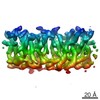 8313MC M: map data used to model this data C: citing same article ( |
|---|---|
| Similar structure data |
-Links
 PDBj
PDBj- Assembly
| Deposited unit | 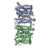
|
|---|---|
| 1 |
|
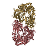
| Symmetry | Helical symmetry: (Circular symmetry: 1 / Dyad axis: yes / N subunits divisor: 1 / Num. of operations: 1 / Rise per n subunits: 4.8 Å / Rotation per n subunits: 37.35 °) |
-Components
| #1: Protein | Mass: 53829.766 Da / Num. of mol.: 2 Source method: isolated from a genetically manipulated source Source: (gene. exp.) |
|---|
-Experimental details
-Experiment
| Experiment | Method: ELECTRON MICROSCOPY |
|---|---|
| EM experiment | Aggregation state: HELICAL ARRAY / 3D reconstruction method: helical reconstruction |
- Sample preparation
| Component | Name: Bor1p dimer in an inward-facing conformation / Type: COMPLEX / Entity ID: all / Source: RECOMBINANT | |||||||||||||||||||||||||
|---|---|---|---|---|---|---|---|---|---|---|---|---|---|---|---|---|---|---|---|---|---|---|---|---|---|---|
| Molecular weight | Value: 0.06 MDa / Experimental value: NO | |||||||||||||||||||||||||
| Source (natural) | Organism: | |||||||||||||||||||||||||
| Source (recombinant) | Organism: | |||||||||||||||||||||||||
| Buffer solution | pH: 7 / Details: Buffer was changed twice per day. | |||||||||||||||||||||||||
| Buffer component |
| |||||||||||||||||||||||||
| Specimen | Conc.: 0.25 mg/ml / Embedding applied: NO / Shadowing applied: NO / Staining applied: NO / Vitrification applied: YES Details: The protein was solubilized in 1% n-Dodecyl beta-D-maltoside and exchanged into heptaethyleneglycol-n-dodecylether (C12E7) while bound to the IgG Sepharose affinity column. | |||||||||||||||||||||||||
| Specimen support | Grid material: COPPER / Grid mesh size: 400 divisions/in. / Grid type: EMS | |||||||||||||||||||||||||
| Vitrification | Instrument: HOMEMADE PLUNGER / Cryogen name: ETHANE / Humidity: 90 % |
- Electron microscopy imaging
| Experimental equipment |  Model: Tecnai F20 / Image courtesy: FEI Company |
|---|---|
| Microscopy | Model: FEI TECNAI F20 |
| Electron gun | Electron source:  FIELD EMISSION GUN / Accelerating voltage: 200 kV / Illumination mode: FLOOD BEAM FIELD EMISSION GUN / Accelerating voltage: 200 kV / Illumination mode: FLOOD BEAM |
| Electron lens | Mode: BRIGHT FIELD / Nominal magnification: 19000 X / Nominal defocus max: 2500 nm / Nominal defocus min: 1000 nm / Calibrated defocus min: 850 nm / Calibrated defocus max: 2700 nm / Cs: 2 mm |
| Specimen holder | Cryogen: NITROGEN Specimen holder model: GATAN 626 SINGLE TILT LIQUID NITROGEN CRYO TRANSFER HOLDER |
| Image recording | Average exposure time: 6.75 sec. / Electron dose: 45 e/Å2 / Detector mode: SUPER-RESOLUTION / Film or detector model: GATAN K2 SUMMIT (4k x 4k) / Num. of grids imaged: 2 / Num. of real images: 252 |
| Image scans | Width: 3710 / Height: 3710 / Movie frames/image: 27 / Used frames/image: 2-27 |
- Processing
| EM software |
| ||||||||||||||||||||||||||||||||||||||||||||||||||
|---|---|---|---|---|---|---|---|---|---|---|---|---|---|---|---|---|---|---|---|---|---|---|---|---|---|---|---|---|---|---|---|---|---|---|---|---|---|---|---|---|---|---|---|---|---|---|---|---|---|---|---|
| CTF correction | Type: PHASE FLIPPING AND AMPLITUDE CORRECTION | ||||||||||||||||||||||||||||||||||||||||||||||||||
| Helical symmerty | Angular rotation/subunit: 37.35 ° / Axial rise/subunit: 4.8 Å / Axial symmetry: C1 | ||||||||||||||||||||||||||||||||||||||||||||||||||
| Particle selection | Num. of particles selected: 87 | ||||||||||||||||||||||||||||||||||||||||||||||||||
| 3D reconstruction | Resolution: 5.9 Å / Resolution method: FSC 0.143 CUT-OFF / Num. of particles: 75 Details: Reconstruction was done using D1 symmetry because of an existing two-fold symmetry in the unit cells composing the helical lattice. This two-fold axis runs perpendicular to the helical axis. ...Details: Reconstruction was done using D1 symmetry because of an existing two-fold symmetry in the unit cells composing the helical lattice. This two-fold axis runs perpendicular to the helical axis. For the final structure, a filter was applied to the map in order to compensate for resolution-dependent amplitude falloff. To do so, we built a model by arranging UraA in a helical assembly in order to mimic the mass distribution in Bor1p tubes. Fourier transforms from this model and from the experimental maps were then rotationally averaged to produce 1D scattering profiles. The resolution-dependent amplitude ratio from these profiles was used as a filter that was applied to the experimental amplitudes using SPARX routines. Finally, a low-pass filter was applied with a 5 Angstrom stop-band frequency. Symmetry type: HELICAL | ||||||||||||||||||||||||||||||||||||||||||||||||||
| Atomic model building | Protocol: FLEXIBLE FIT / Space: REAL / Target criteria: cross-correlation coefficient Details: A homology model was first created using human AE1 (PDB entry 4YZF) as a template using MODELLER, placed into the density map using SITUS, and then fitted into the map using the Molecular ...Details: A homology model was first created using human AE1 (PDB entry 4YZF) as a template using MODELLER, placed into the density map using SITUS, and then fitted into the map using the Molecular Dynamics Flexible Fitting (MDFF) method. | ||||||||||||||||||||||||||||||||||||||||||||||||||
| Atomic model building | 3D fitting-ID: 1 / Accession code: 4YZF / Initial refinement model-ID: 1 / Pdb chain residue range: 418-911 / PDB-ID: 4YZF / Source name: PDB / Type: experimental model
|