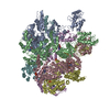
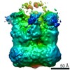
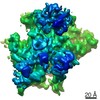
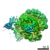
Journal: Sci Adv / Year: 2017 Title: Potyvirus virion structure shows conserved protein fold and RNA binding site in ssRNA viruses. Authors: Miguel Zamora / Eduardo Méndez-López / Xabier Agirrezabala / Rebeca Cuesta / José L Lavín / M Amelia Sánchez-Pina / Miguel A Aranda / Mikel Valle / Abstract: Potyviruses constitute the second largest genus of plant viruses and cause important economic losses in a large variety of crops; however, the atomic structure of their particles remains unknown. ...Potyviruses constitute the second largest genus of plant viruses and cause important economic losses in a large variety of crops; however, the atomic structure of their particles remains unknown. Infective potyvirus virions are long flexuous filaments where coat protein (CP) subunits assemble in helical mode bound to a monopartite positive-sense single-stranded RNA [(+)ssRNA] genome. We present the cryo-electron microscopy (cryoEM) structure of the potyvirus watermelon mosaic virus at a resolution of 4.0 Å. The atomic model shows a conserved fold for the CPs of flexible filamentous plant viruses, including a universally conserved RNA binding pocket, which is a potential target for antiviral compounds. This conserved fold of the CP is widely distributed in eukaryotic viruses and is also shared by nucleoproteins of enveloped viruses with segmented (-)ssRNA (negative-sense ssRNA) genomes, including influenza viruses.
Mass: 31463.412 Da / Num. of mol.: 24 / Source method: isolated from a natural source Details: 57 residues from N-terminus and 17 residues from C-terminus are not present in our pdb model due to the absence of densities for them in the cryo-electron microscopy map as a consequence of ...Details: 57 residues from N-terminus and 17 residues from C-terminus are not present in our pdb model due to the absence of densities for them in the cryo-electron microscopy map as a consequence of being high flexible regions in the protein. Source: (natural) Watermelon mosaic virus / References: UniProt: Q70J31
#2: RNA chain
... RNA (5'-R(P*UP*UP*UP*UP*U)-3')
Mass: 1485.872 Da / Num. of mol.: 24 / Source method: isolated from a natural source / Source: (natural) Watermelon mosaic virus
-
Experimental details
-
Experiment
Experiment
Method: ELECTRON MICROSCOPY
EM experiment
Aggregation state: FILAMENT / 3D reconstruction method: helical reconstruction
In the structure databanks used in Yorodumi, some data are registered as the other names, "COVID-19 virus" and "2019-nCoV". Here are the details of the virus and the list of structure data.
Jan 31, 2019. EMDB accession codes are about to change! (news from PDBe EMDB page)
EMDB accession codes are about to change! (news from PDBe EMDB page)
The allocation of 4 digits for EMDB accession codes will soon come to an end. Whilst these codes will remain in use, new EMDB accession codes will include an additional digit and will expand incrementally as the available range of codes is exhausted. The current 4-digit format prefixed with “EMD-” (i.e. EMD-XXXX) will advance to a 5-digit format (i.e. EMD-XXXXX), and so on. It is currently estimated that the 4-digit codes will be depleted around Spring 2019, at which point the 5-digit format will come into force.
The EM Navigator/Yorodumi systems omit the EMD- prefix.
Related info.:Q: What is EMD? / ID/Accession-code notation in Yorodumi/EM Navigator
Yorodumi is a browser for structure data from EMDB, PDB, SASBDB, etc.
This page is also the successor to EM Navigator detail page, and also detail information page/front-end page for Omokage search.
The word "yorodu" (or yorozu) is an old Japanese word meaning "ten thousand". "mi" (miru) is to see.
Related info.:EMDB / PDB / SASBDB / Comparison of 3 databanks / Yorodumi Search / Aug 31, 2016. New EM Navigator & Yorodumi / Yorodumi Papers / Jmol/JSmol / Function and homology information / Changes in new EM Navigator and Yorodumi
 Movie
Movie Controller


 Open data
 Basic information
 Components
 Keywords
 Function and homology information
 Watermelon mosaic virus
 Authors
 Spain, 1items
  Citation
 Structure visualization
 Downloads & links
 Other downloads
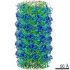


 PDBj
PDBj Assembly
 Sample preparation
 Electron microscopy imaging
 FIELD EMISSION GUN / Accelerating voltage: 300 kV / Illumination mode: FLOOD BEAM
 Processing