 ムービー
ムービー コントローラー


+ データを開く

- 基本情報
基本情報
| 登録情報 | データベース: PDB / ID: 5i08 | ||||||
|---|---|---|---|---|---|---|---|
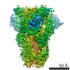
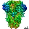
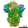
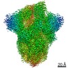
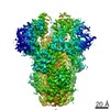
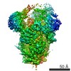
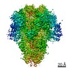
| タイトル | Prefusion structure of a human coronavirus spike protein | ||||||
 要素 要素 | Spike glycoprotein,Foldon chimera | ||||||
 キーワード キーワード | VIRAL PROTEIN / coronavirus / glycoprotein / prefusion | ||||||
| 機能・相同性 |  機能・相同性情報 機能・相同性情報virion component / endocytosis involved in viral entry into host cell / host cell endoplasmic reticulum-Golgi intermediate compartment membrane / receptor-mediated virion attachment to host cell / fusion of virus membrane with host plasma membrane / fusion of virus membrane with host endosome membrane / viral envelope / host cell plasma membrane / virion membrane / identical protein binding / membrane 類似検索 - 分子機能 | ||||||
| 生物種 |  Human coronavirus HKU1 (ウイルス)Enterobacteria phage T4 (ファージ) Human coronavirus HKU1 (ウイルス)Enterobacteria phage T4 (ファージ) | ||||||
| 手法 | 電子顕微鏡法 / 単粒子再構成法 / クライオ電子顕微鏡法 / 解像度: 4.04 Å | ||||||
 データ登録者 データ登録者 | Kirchdoerfer, R.N. / Cottrell, C.A. / Wang, N. / Pallesen, J. / Yassine, H.M. / Turner, H.L. / Corbett, K.S. / Graham, B.S. / McLellan, J.S. / Ward, A.B. | ||||||
 引用 引用 | ジャーナル: Nature / 年: 2016 タイトル: Pre-fusion structure of a human coronavirus spike protein. 著者: Robert N Kirchdoerfer / Christopher A Cottrell / Nianshuang Wang / Jesper Pallesen / Hadi M Yassine / Hannah L Turner / Kizzmekia S Corbett / Barney S Graham / Jason S McLellan / Andrew B Ward /  要旨: HKU1 is a human betacoronavirus that causes mild yet prevalent respiratory disease, and is related to the zoonotic SARS and MERS betacoronaviruses, which have high fatality rates and pandemic ...HKU1 is a human betacoronavirus that causes mild yet prevalent respiratory disease, and is related to the zoonotic SARS and MERS betacoronaviruses, which have high fatality rates and pandemic potential. Cell tropism and host range is determined in part by the coronavirus spike (S) protein, which binds cellular receptors and mediates membrane fusion. As the largest known class I fusion protein, its size and extensive glycosylation have hindered structural studies of the full ectodomain, thus preventing a molecular understanding of its function and limiting development of effective interventions. Here we present the 4.0 Å resolution structure of the trimeric HKU1 S protein determined using single-particle cryo-electron microscopy. In the pre-fusion conformation, the receptor-binding subunits, S1, rest above the fusion-mediating subunits, S2, preventing their conformational rearrangement. Surprisingly, the S1 C-terminal domains are interdigitated and form extensive quaternary interactions that occlude surfaces known in other coronaviruses to bind protein receptors. These features, along with the location of the two protease sites known to be important for coronavirus entry, provide a structural basis to support a model of membrane fusion mediated by progressive S protein destabilization through receptor binding and proteolytic cleavage. These studies should also serve as a foundation for the structure-based design of betacoronavirus vaccine immunogens. | ||||||
| 履歴 |
|
- 構造の表示
構造の表示
| ムービー |
ムービービューア |
|---|---|
| 構造ビューア | 分子: MolmilJmol/JSmol |
- ダウンロードとリンク
-ダウンロード
| PDBx/mmCIF形式 | 5i08.cif.gz | 519.6 KB | 表示 | PDBx/mmCIF形式 |
|---|---|---|---|---|
| PDB形式 | pdb5i08.ent.gz | 414.1 KB | 表示 | PDB形式 |
| PDBx/mmJSON形式 | 5i08.json.gz | ツリー表示 | PDBx/mmJSON形式 | |
| その他 |  その他のダウンロード その他のダウンロード |
-検証レポート
| 文書・要旨 | 5i08_validation.pdf.gz | 1.2 MB | 表示 | wwPDB検証レポート |
|---|---|---|---|---|
| 文書・詳細版 | 5i08_full_validation.pdf.gz | 1.2 MB | 表示 | |
| XML形式データ | 5i08_validation.xml.gz | 80.9 KB | 表示 | |
| CIF形式データ | 5i08_validation.cif.gz | 123.8 KB | 表示 | |
| アーカイブディレクトリ | https://data.pdbj.org/pub/pdb/validation_reports/i0/5i08ftp://data.pdbj.org/pub/pdb/validation_reports/i0/5i08 | HTTPS FTP |
-関連構造データ
-リンク
 PDBj
PDBj

- 集合体
集合体
| 登録構造単位 | 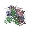
|
|---|---|
| 1 |
|
-要素
| #1: タンパク質 | 分子量: 144295.891 Da / 分子数: 3 変異: R751G, R752G, K753S, R754G, R755S,R751G, R752G, K753S, R754G, R755S 由来タイプ: 組換発現 由来: (組換発現) Human coronavirus HKU1 (isolate N5) (ウイルス), (組換発現) Enterobacteria phage T4 (ファージ)株: isolate N5 / 遺伝子: S, 3, wac / プラスミド: pVRC-8400 / 細胞株 (発現宿主): FreeStyle 293F / 発現宿主:  Homo sapiens (ヒト) / 参照: UniProt: Q0ZME7, UniProt: P10104 Homo sapiens (ヒト) / 参照: UniProt: Q0ZME7, UniProt: P10104 |
|---|
-実験情報
-実験
| 実験 | 手法: 電子顕微鏡法 |
|---|---|
| EM実験 | 試料の集合状態: PARTICLE / 3次元再構成法: 単粒子再構成法 |
- 試料調製
試料調製
| 構成要素 | 名称: HKU1 spike with attached foldon domain and mutated furin-cleavage site タイプ: COMPLEX / Entity ID: all / 由来: RECOMBINANT | |||||||||||||||
|---|---|---|---|---|---|---|---|---|---|---|---|---|---|---|---|---|
| 分子量 | 値: 0.42 MDa / 実験値: NO | |||||||||||||||
| 由来(天然) | 生物種: Human coronavirus HKU1 (isolate N5) (ウイルス) | |||||||||||||||
| 由来(組換発現) | 生物種: Homo sapiens (ヒト) / 細胞: FreeStyle 293F / プラスミド: pVRC8400 | |||||||||||||||
| 緩衝液 | pH: 8 | |||||||||||||||
| 緩衝液成分 |
| |||||||||||||||
| 試料 | 濃度: 0.27 mg/ml / 包埋: NO / シャドウイング: NO / 染色: NO / 凍結: YES | |||||||||||||||
| 染色 | タイプ: NEGATIVE 詳細: 3 uL sample was applied to grid for 30 seconds and then blotted. Grids were stained with 3 uL 1% uranyl formate for 60 seconds followed by blotting. 染色剤: uranyl formate | |||||||||||||||
| 試料支持 | グリッドの材料: COPPER / グリッドのサイズ: 400 divisions/in. / グリッドのタイプ: EMS CF-2/2-4C C-Flat | |||||||||||||||
| 急速凍結 | 装置: HOMEMADE PLUNGER / 凍結剤: ETHANE 詳細: 3 uL sample was applied to grid, blotted, and plunged into liquid ethane. |
- 電子顕微鏡撮影
| 実験機器 |  モデル: Titan Krios / 画像提供: FEI Company |
|---|---|
| 顕微鏡 | モデル: FEI TITAN KRIOS |
| 電子銃 | 電子線源:  FIELD EMISSION GUN / 加速電圧: 300 kV / 照射モード: FLOOD BEAM FIELD EMISSION GUN / 加速電圧: 300 kV / 照射モード: FLOOD BEAM |
| 電子レンズ | モード: BRIGHT FIELD / 倍率(公称値): 22500 X / 倍率(補正後): 22500 X / 最大 デフォーカス(公称値): 3500 nm / 最小 デフォーカス(公称値): 1000 nm |
| 試料ホルダ | 凍結剤: NITROGEN 試料ホルダーモデル: FEI TITAN KRIOS AUTOGRID HOLDER |
| 撮影 | 平均露光時間: 10 sec. / 電子線照射量: 57 e/Å2 / 検出モード: COUNTING フィルム・検出器のモデル: GATAN K2 SUMMIT (4k x 4k) 撮影したグリッド数: 1 / 実像数: 1049 詳細: Images were collected using Legionon and processed using Appion. |
| 画像スキャン | 動画フレーム数/画像: 50 |
- 解析
解析
| EMソフトウェア |
| |||||||||||||||||||||||||||||||||||||||||||||||||||||||||||||||||
|---|---|---|---|---|---|---|---|---|---|---|---|---|---|---|---|---|---|---|---|---|---|---|---|---|---|---|---|---|---|---|---|---|---|---|---|---|---|---|---|---|---|---|---|---|---|---|---|---|---|---|---|---|---|---|---|---|---|---|---|---|---|---|---|---|---|---|
| CTF補正 | タイプ: PHASE FLIPPING AND AMPLITUDE CORRECTION | |||||||||||||||||||||||||||||||||||||||||||||||||||||||||||||||||
| 粒子像の選択 | 選択した粒子像数: 39164 詳細: 2188 particles were selected from a subset of the data using DoG Picker. These particles were used to generate a 3D model from which back projections were derived. Back projection images were ...詳細: 2188 particles were selected from a subset of the data using DoG Picker. These particles were used to generate a 3D model from which back projections were derived. Back projection images were used as templates for picking particles from the entire dataset using FindEM. | |||||||||||||||||||||||||||||||||||||||||||||||||||||||||||||||||
| 対称性 | 点対称性: C3 (3回回転対称) | |||||||||||||||||||||||||||||||||||||||||||||||||||||||||||||||||
| 3次元再構成 | 解像度: 4.04 Å / 解像度の算出法: FSC 0.143 CUT-OFF / 粒子像の数: 31435 / アルゴリズム: BACK PROJECTION / 対称性のタイプ: POINT | |||||||||||||||||||||||||||||||||||||||||||||||||||||||||||||||||
| 原子モデル構築 | B value: 117 / プロトコル: AB INITIO MODEL / 空間: REAL / Target criteria: EMRinger 詳細: Model building and refinement were conducted using a combination of software programs. |
