 ムービー
ムービー コントローラー


+ データを開く

- 基本情報
基本情報
| 登録情報 | データベース: PDB / ID: 5abb | ||||||
|---|---|---|---|---|---|---|---|
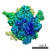
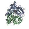
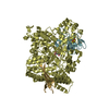
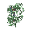
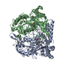
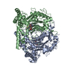
| タイトル | Visualization of a polytopic membrane protein during SecY-mediated membrane insertion | ||||||
 要素 要素 |
| ||||||
 キーワード キーワード | TRANSLATION / RIBOSOME / MEMBRANE PROTEIN / TRANSLOCON | ||||||
| 機能・相同性 |  機能・相同性情報 機能・相同性情報light-activated monoatomic ion channel activity / cell envelope Sec protein transport complex / protein transport by the Sec complex / intracellular protein transmembrane transport / SRP-dependent cotranslational protein targeting to membrane, translocation / signal sequence receptor activity / Secretion of toxins / photoreceptor activity / transmembrane protein transporter activity / protein secretion ...light-activated monoatomic ion channel activity / cell envelope Sec protein transport complex / protein transport by the Sec complex / intracellular protein transmembrane transport / SRP-dependent cotranslational protein targeting to membrane, translocation / signal sequence receptor activity / Secretion of toxins / photoreceptor activity / transmembrane protein transporter activity / protein secretion / phototransduction / protein targeting / proton transmembrane transport / intracellular protein transport / membrane / plasma membrane 類似検索 - 分子機能 | ||||||
| 生物種 |  | ||||||
| 手法 | 電子顕微鏡法 / 単粒子再構成法 / クライオ電子顕微鏡法 / 解像度: 8 Å | ||||||
 データ登録者 データ登録者 | Bischoff, L. / Wickles, S. / Berninghausen, O. / vanderSluis, E. / Beckmann, R. | ||||||
 引用 引用 | ジャーナル: Nat Commun / 年: 2014 タイトル: Visualization of a polytopic membrane protein during SecY-mediated membrane insertion. 著者: Lukas Bischoff / Stephan Wickles / Otto Berninghausen / Eli O van der Sluis / Roland Beckmann /  要旨: The biogenesis of polytopic membrane proteins occurs co-translationally on ribosomes that are tightly bound to a membrane-embedded protein-conducting channel: the Sec-complex. The path that is ...The biogenesis of polytopic membrane proteins occurs co-translationally on ribosomes that are tightly bound to a membrane-embedded protein-conducting channel: the Sec-complex. The path that is followed by nascent proteins inside the ribosome and the Sec-complex is relatively well established; however, it is not clear what the fate of the N-terminal transmembrane domains (TMDs) of polytopic membrane proteins is when the C-terminal TMDs domains are not yet synthesized. Here, we present the sub-nanometer cryo-electron microscopy structure of an in vivo generated ribosome-SecY complex that carries a membrane insertion intermediate of proteorhodopsin (PR). The structure reveals a pre-opened Sec-complex and the first two TMDs of PR already outside the SecY complex directly in front of its proposed lateral gate. Thus, our structure is in agreement with positioning of N-terminal TMDs at the periphery of SecY, and in addition, it provides clues for the molecular mechanism underlying membrane protein topogenesis. | ||||||
| 履歴 |
|
- 構造の表示
構造の表示
| ムービー |
ムービービューア |
|---|---|
| 構造ビューア | 分子: MolmilJmol/JSmol |
- ダウンロードとリンク
-ダウンロード
| PDBx/mmCIF形式 | 5abb.cif.gz | 90.6 KB | 表示 | PDBx/mmCIF形式 |
|---|---|---|---|---|
| PDB形式 | pdb5abb.ent.gz | 54.9 KB | 表示 | PDB形式 |
| PDBx/mmJSON形式 | 5abb.json.gz | ツリー表示 | PDBx/mmJSON形式 | |
| その他 |  その他のダウンロード その他のダウンロード |
-検証レポート
| アーカイブディレクトリ | https://data.pdbj.org/pub/pdb/validation_reports/ab/5abbftp://data.pdbj.org/pub/pdb/validation_reports/ab/5abb | HTTPS FTP |
|---|
-関連構造データ


-リンク
 PDBj
PDBj


- 集合体
集合体
| 登録構造単位 | 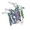
|
|---|---|
| 1 |
|
-要素
| #1: タンパク質 | 分子量: 48553.375 Da / 分子数: 1 / 由来タイプ: 天然 / 由来: (天然) |
|---|---|
| #2: タンパク質 | 分子量: 12623.296 Da / 分子数: 1 / Fragment: UNP RESIDUES 12-127 / 由来タイプ: 天然 / 由来: (天然) |
| #3: タンパク質 | 分子量: 7634.684 Da / 分子数: 1 / Fragment: UNP RESIDUES 19-83 / 由来タイプ: 天然 / 由来: (天然) |
-実験情報
-実験
| 実験 | 手法: 電子顕微鏡法 |
|---|---|
| EM実験 | 試料の集合状態: PARTICLE / 3次元再構成法: 単粒子再構成法 |
- 試料調製
試料調製
| 構成要素 | 名称: TnaC stalled E.coli ribosome in complex with SecYE / タイプ: RIBOSOME |
|---|---|
| 緩衝液 | 名称: 20 MM TRIS 150 MM NH4CL 10 MM MGCL2 0.05%DDM 125 MM SUCROSE pH: 7.5 詳細: 20 MM TRIS 150 MM NH4CL 10 MM MGCL2 0.05%DDM 125 MM SUCROSE |
| 試料 | 包埋: NO / シャドウイング: NO / 染色: NO / 凍結: YES |
| 試料支持 | 詳細: HOLEY CARBON |
| 急速凍結 | 装置: FEI VITROBOT MARK IV / 凍結剤: ETHANE |
- 電子顕微鏡撮影
| 実験機器 |  モデル: Titan Krios / 画像提供: FEI Company |
|---|---|
| 顕微鏡 | モデル: FEI TITAN KRIOS / 日付: 2012年5月21日 |
| 電子銃 | 電子線源:  FIELD EMISSION GUN / 加速電圧: 200 kV / 照射モード: SPOT SCAN FIELD EMISSION GUN / 加速電圧: 200 kV / 照射モード: SPOT SCAN |
| 電子レンズ | モード: BRIGHT FIELD / 倍率(公称値): 148721 X / 最大 デフォーカス(公称値): 3500 nm / 最小 デフォーカス(公称値): 1000 nm |
| 撮影 | 電子線照射量: 20 e/Å2 フィルム・検出器のモデル: TVIPS TEMCAM-F416 (4k x 4k) |
| 放射波長 | 相対比: 1 |
- 解析
解析
| EMソフトウェア |
| ||||||||||||
|---|---|---|---|---|---|---|---|---|---|---|---|---|---|
| 対称性 | 点対称性: C1 (非対称) | ||||||||||||
| 3次元再構成 | 手法: REAL / 解像度: 8 Å / 粒子像の数: 47471 / 対称性のタイプ: POINT | ||||||||||||
| 精密化 | 最高解像度: 8 Å | ||||||||||||
| 精密化ステップ | サイクル: LAST / 最高解像度: 8 Å
|
