protein localization to paranode region of axon / cytoskeletal protein-membrane anchor activity / paranodal junction assembly / protein localization to juxtaparanode region of axon / myelin maintenance / paranode region of axon / cortical cytoskeleton organization / juxtaparanode region of axon / actomyosin structure organization / cortical actin cytoskeleton organization ...protein localization to paranode region of axon / cytoskeletal protein-membrane anchor activity / paranodal junction assembly / protein localization to juxtaparanode region of axon / myelin maintenance / paranode region of axon / cortical cytoskeleton organization / juxtaparanode region of axon / actomyosin structure organization / cortical actin cytoskeleton organization / Sensory processing of sound by outer hair cells of the cochlea / Neurexins and neuroligins / Sensory processing of sound by inner hair cells of the cochlea / regulation of neurotransmitter receptor localization to postsynaptic specialization membrane / neuron projection morphogenesis / protein localization to plasma membrane / structural constituent of cytoskeleton / cell-cell junction / cell junction / regulation of cell shape / actin binding / cytoskeleton / postsynapse / cilium / ciliary basal body / apoptotic process / glutamatergic synapse / plasma membrane / cytosol Similarity search - Function
Band 4.1-like protein 3 / SAB domain / Band 4.1, C-terminal / SAB domain / 4.1 protein C-terminal domain (CTD) / FERM adjacent (FA) / FERM adjacent (FA) / FERM adjacent (FA) / Ezrin/radixin/moesin-like / FERM, C-terminal PH-like domain ...Band 4.1-like protein 3 / SAB domain / Band 4.1, C-terminal / SAB domain / 4.1 protein C-terminal domain (CTD) / FERM adjacent (FA) / FERM adjacent (FA) / FERM adjacent (FA) / Ezrin/radixin/moesin-like / FERM, C-terminal PH-like domain / FERM C-terminal PH-like domain / FERM C-terminal PH-like domain / FERM, N-terminal / FERM N-terminal domain / FERM domain signature 1. / FERM conserved site / FERM domain signature 2. / FERM central domain / FERM/acyl-CoA-binding protein superfamily / FERM central domain / FERM superfamily, second domain / FERM domain / FERM domain profile. / Band 4.1 domain / Band 4.1 homologues / PH-like domain superfamily / Ubiquitin-like domain superfamily Similarity search - Domain/homology
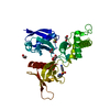
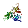
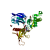
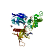
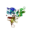
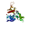
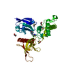
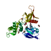
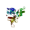
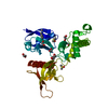
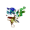
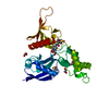
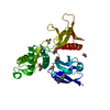
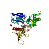
The FERM domain of HUMAN EPB41L3 screened against the Enamine DSI-poised fragment library by X-ray Crystallography at the XChem facility of Diamond Light Source beamline I04-1
Isoform2ofBand4.1-likeprotein3 / 4.1B / Differentially expressed in adenocarcinoma of the lung protein 1 / DAL-1 / Erythrocyte ...4.1B / Differentially expressed in adenocarcinoma of the lung protein 1 / DAL-1 / Erythrocyte membrane protein band 4.1-like 3
Mass: 33395.305 Da / Num. of mol.: 1 Source method: isolated from a genetically manipulated source Source: (gene. exp.) Homo sapiens (human) / Gene: EPB41L3, DAL1, KIAA0987 / Production host: Escherichia coli (E. coli) / References: UniProt: Q9Y2J2
In the structure databanks used in Yorodumi, some data are registered as the other names, "COVID-19 virus" and "2019-nCoV". Here are the details of the virus and the list of structure data.
Jan 31, 2019. EMDB accession codes are about to change! (news from PDBe EMDB page)
EMDB accession codes are about to change! (news from PDBe EMDB page)
The allocation of 4 digits for EMDB accession codes will soon come to an end. Whilst these codes will remain in use, new EMDB accession codes will include an additional digit and will expand incrementally as the available range of codes is exhausted. The current 4-digit format prefixed with “EMD-” (i.e. EMD-XXXX) will advance to a 5-digit format (i.e. EMD-XXXXX), and so on. It is currently estimated that the 4-digit codes will be depleted around Spring 2019, at which point the 5-digit format will come into force.
The EM Navigator/Yorodumi systems omit the EMD- prefix.
Related info.:Q: What is EMD? / ID/Accession-code notation in Yorodumi/EM Navigator
Yorodumi is a browser for structure data from EMDB, PDB, SASBDB, etc.
This page is also the successor to EM Navigator detail page, and also detail information page/front-end page for Omokage search.
The word "yorodu" (or yorozu) is an old Japanese word meaning "ten thousand". "mi" (miru) is to see.
Related info.:EMDB / PDB / SASBDB / Comparison of 3 databanks / Yorodumi Search / Aug 31, 2016. New EM Navigator & Yorodumi / Yorodumi Papers / Jmol/JSmol / Function and homology information / Changes in new EM Navigator and Yorodumi
 Movie
Movie Controller

 Yorodumi
 Open data
 Basic information
 Components
 Keywords
 Function and homology information
 Homo sapiens (human)
 X-RAY DIFFRACTION /
  Authors
 Citation
 Structure visualization
 Downloads & links
 Other downloads


 PDBj
PDBj


 Assembly
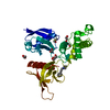

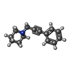
 Mass: 199.291 Da / Num. of mol.: 1 / Source method: obtained synthetically / Formula: C14H17N / Feature type: SUBJECT OF INVESTIGATION
 Mass: 78.133 Da / Num. of mol.: 1 / Source method: obtained synthetically / Formula: C2H6OS / Comment: DMSO, precipitant*YM
 Mass: 62.068 Da / Num. of mol.: 6 / Source method: obtained synthetically / Formula: C2H6O2
 Mass: 18.015 Da / Num. of mol.: 151 / Source method: isolated from a natural source / Formula: H2O
 Sample preparation
 / Beamline: I04-1 / Wavelength: 0.91587 Å
 Processing