 Movie
Movie Controller


+ Open data

- Basic information
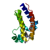
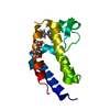
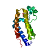
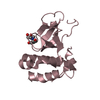
| Entry | Database: PDB / ID: 5nr3 | ||||||
|---|---|---|---|---|---|---|---|
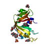
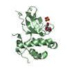
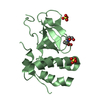
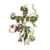
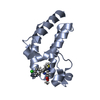
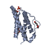
| Title | Human DNMT3B PWWP domain in complex with ethambutol | ||||||
 Components Components | DNA (cytosine-5)-methyltransferase 3B | ||||||
 Keywords Keywords | TRANSFERASE / DNMT3B PWWP DOMAIN / HISTONE BINDING / BETA BARREL / LIGAND | ||||||
| Function / homology |  Function and homology information Function and homology informationDNA (cytosine-5-)-methyltransferase activity, acting on CpG substrates / DNA-methyltransferase activity / DNA (cytosine-5-)-methyltransferase / DNA (cytosine-5-)-methyltransferase activity / SUMOylation of DNA methylation proteins / catalytic complex / DNA methylation / PRC2 methylates histones and DNA / Defective pyroptosis / NoRC negatively regulates rRNA expression ...DNA (cytosine-5-)-methyltransferase activity, acting on CpG substrates / DNA-methyltransferase activity / DNA (cytosine-5-)-methyltransferase / DNA (cytosine-5-)-methyltransferase activity / SUMOylation of DNA methylation proteins / catalytic complex / DNA methylation / PRC2 methylates histones and DNA / Defective pyroptosis / NoRC negatively regulates rRNA expression / transcription corepressor activity / methylation / positive regulation of gene expression / negative regulation of transcription by RNA polymerase II / DNA binding / zinc ion binding / nucleoplasm / nucleus Similarity search - Function | ||||||
| Biological species |  Homo sapiens (human) Homo sapiens (human) | ||||||
| Method |  X-RAY DIFFRACTION / SYNCHROTRON / MOLECULAR REPLACEMENT / molecular replacement / Resolution: 2.3 Å X-RAY DIFFRACTION / SYNCHROTRON / MOLECULAR REPLACEMENT / molecular replacement / Resolution: 2.3 Å | ||||||
 Authors Authors | Rondelet, G. / Wouters, J. | ||||||
| Funding support |  Belgium, 1items Belgium, 1items
| ||||||
 Citation Citation | Journal: To be published Title: Targeting PWWP domain of DNA methyltransferase 3B for epigenetic cancer therapy: Identification and structural characterization of new potential protein-protein interaction inhibitors Authors: Rondelet, G. / Dal Maso, T. / Maniquet, A. / Themans, Q. / Wouters, J. | ||||||
| History |
|
- Structure visualization
| Structure viewer | Molecule: MolmilJmol/JSmol |
|---|
- Downloads & links
-Download
| PDBx/mmCIF format | 5nr3.cif.gz | 71 KB | Display | PDBx/mmCIF format |
|---|---|---|---|---|
| PDB format | pdb5nr3.ent.gz | 52.1 KB | Display | PDB format |
| PDBx/mmJSON format | 5nr3.json.gz | Tree view | PDBx/mmJSON format | |
| Others |  Other downloads Other downloads |
-Validation report
| Arichive directory | https://data.pdbj.org/pub/pdb/validation_reports/nr/5nr3ftp://data.pdbj.org/pub/pdb/validation_reports/nr/5nr3 | HTTPS FTP |
|---|
-Related structure data
| Related structure data |  5nrrC  5nrsC  5nrvC  5nv0C  5nv2C  5nv7C  5nvoC 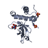 3qkjS S: Starting model for refinement C: citing same article ( |
|---|---|
| Similar structure data |
-Links
 PDBj
PDBj


- Assembly
| Deposited unit | 
| ||||||||
|---|---|---|---|---|---|---|---|---|---|
| 1 | 
| ||||||||
| 2 | 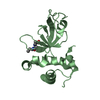
| ||||||||
| Unit cell |
|
-Components
| #1: Protein | Mass: 16698.844 Da / Num. of mol.: 2 / Fragment: UNP residues 218-367 Source method: isolated from a genetically manipulated source Source: (gene. exp.) Homo sapiens (human) / Gene: DNMT3B / Plasmid: pET28-MHL / Production host:  References: UniProt: Q9UBC3, DNA (cytosine-5-)-methyltransferase #2: Chemical |   Mass: 204.310 Da / Num. of mol.: 2 / Source method: obtained synthetically / Formula: C10H24N2O2 / Comment: medication*YM Mass: 204.310 Da / Num. of mol.: 2 / Source method: obtained synthetically / Formula: C10H24N2O2 / Comment: medication*YM#3: Chemical |   Mass: 96.063 Da / Num. of mol.: 3 / Source method: obtained synthetically / Formula: SO4 Mass: 96.063 Da / Num. of mol.: 3 / Source method: obtained synthetically / Formula: SO4#4: Water | ChemComp-HOH / |  Mass: 18.015 Da / Num. of mol.: 171 / Source method: isolated from a natural source / Formula: H2O Mass: 18.015 Da / Num. of mol.: 171 / Source method: isolated from a natural source / Formula: H2O |
|---|
-Experimental details
-Experiment
| Experiment | Method: X-RAY DIFFRACTION / Number of used crystals: 1 |
|---|
- Sample preparation
| Crystal | Density Matthews: 3.71 Å3/Da / Density % sol: 66.82 % |
|---|---|
| Crystal grow | Temperature: 298 K / Method: vapor diffusion, sitting drop / Details: 0.1 M MES,0.2 M Li2SO4, 23-33% PEG 3350 / PH range: 5.7-6.7 |
-Data collection
| Diffraction | Mean temperature: 100 K |
|---|---|
| Diffraction source | Source: SYNCHROTRON / Site: SOLEIL  / Beamline: PROXIMA 2 / Wavelength: 0.9801 Å / Beamline: PROXIMA 2 / Wavelength: 0.9801 Å |
| Detector | Type: ADSC QUANTUM 315r / Detector: CCD / Date: Sep 19, 2015 / Details: X-RAY FLUORESCCENCE DETECTOR |
| Radiation | Monochromator: CRYOGENICALLY COOLED CHANNEL-CUT SI[111] / Protocol: SINGLE WAVELENGTH / Monochromatic (M) / Laue (L): M / Scattering type: x-ray |
| Radiation wavelength | Wavelength: 0.9801 Å / Relative weight: 1 |
| Reflection | Resolution: 2.3→40.391 Å / Num. obs: 22766 / % possible obs: 99.7 % / Redundancy: 9.6 % / Biso Wilson estimate: 40.66 Å2 / CC1/2: 0.999 / Rmerge(I) obs: 0.078 / Net I/σ(I): 27 |
| Reflection shell | Resolution: 2.3→2.43 Å / Redundancy: 9.4 % / Rmerge(I) obs: 0.873 / Mean I/σ(I) obs: 3.87 / Num. unique obs: 3549 / CC1/2: 0.883 / % possible all: 98.6 |
-Phasing
| Phasing | Method: molecular replacement |
|---|
- Processing
| Software |
| ||||||||||||||||||||||||||||||||||||||||||||||||||||||
|---|---|---|---|---|---|---|---|---|---|---|---|---|---|---|---|---|---|---|---|---|---|---|---|---|---|---|---|---|---|---|---|---|---|---|---|---|---|---|---|---|---|---|---|---|---|---|---|---|---|---|---|---|---|---|---|
| Refinement | Method to determine structure: MOLECULAR REPLACEMENT Starting model: 3QKJ Resolution: 2.3→40.391 Å / SU ML: 0.25 / Cross valid method: FREE R-VALUE / σ(F): 1.36 / Phase error: 24.44
| ||||||||||||||||||||||||||||||||||||||||||||||||||||||
| Solvent computation | Shrinkage radii: 0.9 Å / VDW probe radii: 1.11 Å | ||||||||||||||||||||||||||||||||||||||||||||||||||||||
| Displacement parameters | Biso max: 88.12 Å2 / Biso mean: 35.93 Å2 / Biso min: 13.85 Å2 | ||||||||||||||||||||||||||||||||||||||||||||||||||||||
| Refinement step | Cycle: final / Resolution: 2.3→40.391 Å
| ||||||||||||||||||||||||||||||||||||||||||||||||||||||
| Refine LS restraints |
| ||||||||||||||||||||||||||||||||||||||||||||||||||||||
| LS refinement shell | Refine-ID: X-RAY DIFFRACTION / Rfactor Rfree error: 0 / Total num. of bins used: 8 / % reflection obs: 100 %
|
