 Movie
Movie Controller


+ Open data

- Basic information
| Entry | Database: PDB / ID: 5lxd | ||||||
|---|---|---|---|---|---|---|---|
| Title | Crystal structure of DYRK2 in complex with EHT 1610 (compound 2) | ||||||
 Components Components | Dual specificity tyrosine-phosphorylation-regulated kinase 2 | ||||||
 Keywords Keywords | TRANSFERASE / kinase / inhibitor / unusual binding mode / Structural Genomics / Structural Genomics Consortium / SGC | ||||||
| Function / homology |  Function and homology information Function and homology informationdual-specificity kinase / negative regulation of calcineurin-NFAT signaling cascade / smoothened signaling pathway / intrinsic apoptotic signaling pathway in response to DNA damage by p53 class mediator / positive regulation of glycogen biosynthetic process / ubiquitin ligase complex / protein serine/threonine/tyrosine kinase activity / regulation of signal transduction by p53 class mediator / manganese ion binding / protein tyrosine kinase activity ...dual-specificity kinase / negative regulation of calcineurin-NFAT signaling cascade / smoothened signaling pathway / intrinsic apoptotic signaling pathway in response to DNA damage by p53 class mediator / positive regulation of glycogen biosynthetic process / ubiquitin ligase complex / protein serine/threonine/tyrosine kinase activity / regulation of signal transduction by p53 class mediator / manganese ion binding / protein tyrosine kinase activity / Regulation of TP53 Activity through Phosphorylation / cytoskeleton / protein phosphorylation / ribonucleoprotein complex / protein serine kinase activity / protein serine/threonine kinase activity / DNA damage response / magnesium ion binding / nucleoplasm / ATP binding / nucleus / cytosol / cytoplasm Similarity search - Function | ||||||
| Biological species |  Homo sapiens (human) Homo sapiens (human) | ||||||
| Method |  X-RAY DIFFRACTION / SYNCHROTRON / MOLECULAR REPLACEMENT / Resolution: 2.58 Å X-RAY DIFFRACTION / SYNCHROTRON / MOLECULAR REPLACEMENT / Resolution: 2.58 Å | ||||||
 Authors Authors | Chaikuad, A. / von Delft, F. / Arrowsmith, C.H. / Edwards, A. / Bountra, C. / Besson, T. / Knapp, S. / Structural Genomics Consortium (SGC) | ||||||
 Citation Citation | Journal: J. Med. Chem. / Year: 2016 Title: An Unusual Binding Model of the Methyl 9-Anilinothiazolo[5,4-f] quinazoline-2-carbimidates (EHT 1610 and EHT 5372) Confers High Selectivity for Dual-Specificity Tyrosine Phosphorylation-Regulated Kinases. Authors: Chaikuad, A. / Diharce, J. / Schroder, M. / Foucourt, A. / Leblond, B. / Casagrande, A.S. / Desire, L. / Bonnet, P. / Knapp, S. / Besson, T. | ||||||
| History |
|
- Structure visualization
| Structure viewer | Molecule: MolmilJmol/JSmol |
|---|
- Downloads & links
-Download
| PDBx/mmCIF format | 5lxd.cif.gz | 335 KB | Display | PDBx/mmCIF format |
|---|---|---|---|---|
| PDB format | pdb5lxd.ent.gz | 274.3 KB | Display | PDB format |
| PDBx/mmJSON format | 5lxd.json.gz | Tree view | PDBx/mmJSON format | |
| Others |  Other downloads Other downloads |
-Validation report
| Arichive directory | https://data.pdbj.org/pub/pdb/validation_reports/lx/5lxdftp://data.pdbj.org/pub/pdb/validation_reports/lx/5lxd | HTTPS FTP |
|---|
-Related structure data


-Links
 PDBj
PDBj- Assembly
| Deposited unit | 
| |||||||||||||||||||||||||||||||||||||||||||||||||||||||||||||||||||||||||||||||||||||||||||||||||||||||||||||||||||||||||||||||||||||||||||||||||||||||||||||||||||||||||||||||||||||||||||||
|---|---|---|---|---|---|---|---|---|---|---|---|---|---|---|---|---|---|---|---|---|---|---|---|---|---|---|---|---|---|---|---|---|---|---|---|---|---|---|---|---|---|---|---|---|---|---|---|---|---|---|---|---|---|---|---|---|---|---|---|---|---|---|---|---|---|---|---|---|---|---|---|---|---|---|---|---|---|---|---|---|---|---|---|---|---|---|---|---|---|---|---|---|---|---|---|---|---|---|---|---|---|---|---|---|---|---|---|---|---|---|---|---|---|---|---|---|---|---|---|---|---|---|---|---|---|---|---|---|---|---|---|---|---|---|---|---|---|---|---|---|---|---|---|---|---|---|---|---|---|---|---|---|---|---|---|---|---|---|---|---|---|---|---|---|---|---|---|---|---|---|---|---|---|---|---|---|---|---|---|---|---|---|---|---|---|---|---|---|---|---|
| 1 | 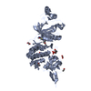
| |||||||||||||||||||||||||||||||||||||||||||||||||||||||||||||||||||||||||||||||||||||||||||||||||||||||||||||||||||||||||||||||||||||||||||||||||||||||||||||||||||||||||||||||||||||||||||||
| 2 | 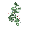
| |||||||||||||||||||||||||||||||||||||||||||||||||||||||||||||||||||||||||||||||||||||||||||||||||||||||||||||||||||||||||||||||||||||||||||||||||||||||||||||||||||||||||||||||||||||||||||||
| Unit cell |
| |||||||||||||||||||||||||||||||||||||||||||||||||||||||||||||||||||||||||||||||||||||||||||||||||||||||||||||||||||||||||||||||||||||||||||||||||||||||||||||||||||||||||||||||||||||||||||||
| Noncrystallographic symmetry (NCS) | NCS domain:
NCS domain segments: Refine code: 2
NCS ensembles :
NCS oper:
|
-Components
| #1: Protein | Mass: 46969.293 Da / Num. of mol.: 2 Source method: isolated from a genetically manipulated source Source: (gene. exp.) Homo sapiens (human) / Gene: DYRK2 / Plasmid: pNIC28-Bsa4 / Production host:  #2: Chemical |   Mass: 383.399 Da / Num. of mol.: 2 / Source method: obtained synthetically / Formula: C18H14FN5O2S Mass: 383.399 Da / Num. of mol.: 2 / Source method: obtained synthetically / Formula: C18H14FN5O2S#3: Chemical | ChemComp-EDO /   Mass: 62.068 Da / Num. of mol.: 10 / Source method: obtained synthetically / Formula: C2H6O2 Mass: 62.068 Da / Num. of mol.: 10 / Source method: obtained synthetically / Formula: C2H6O2#4: Water | ChemComp-HOH / |  Mass: 18.015 Da / Num. of mol.: 193 / Source method: isolated from a natural source / Formula: H2O Mass: 18.015 Da / Num. of mol.: 193 / Source method: isolated from a natural source / Formula: H2OHas protein modification | Y | |
|---|
-Experimental details
-Experiment
| Experiment | Method: X-RAY DIFFRACTION / Number of used crystals: 1 |
|---|
- Sample preparation
| Crystal | Density Matthews: 3.12 Å3/Da / Density % sol: 60.63 % |
|---|---|
| Crystal grow | Temperature: 277.15 K / Method: vapor diffusion, sitting drop / pH: 7.5 / Details: 1.5 M Li2SO4 and 0.1 M HEPES, pH 7.5 |
-Data collection
| Diffraction | Mean temperature: 100 K |
|---|---|
| Diffraction source | Source: SYNCHROTRON / Site: Diamond  / Beamline: I04-1 / Wavelength: 0.92 Å / Beamline: I04-1 / Wavelength: 0.92 Å |
| Detector | Type: DECTRIS PILATUS 2M / Detector: PIXEL / Date: Dec 19, 2012 |
| Radiation | Protocol: SINGLE WAVELENGTH / Monochromatic (M) / Laue (L): M / Scattering type: x-ray |
| Radiation wavelength | Wavelength: 0.92 Å / Relative weight: 1 |
| Reflection | Resolution: 2.58→47.04 Å / Num. obs: 35477 / % possible obs: 97.8 % / Redundancy: 3.4 % / Biso Wilson estimate: 44.6 Å2 / Rmerge(I) obs: 0.091 / Net I/σ(I): 9.1 |
| Reflection shell | Resolution: 2.58→2.72 Å / Redundancy: 3.5 % / Rmerge(I) obs: 0.649 / Mean I/σ(I) obs: 2 / % possible all: 99.4 |
- Processing
| Software |
| ||||||||||||||||||||||||||||||||||||||||||||||||||||||||||||||||||||||||||||||||||||||||||||||||||||||||||||||||||||||||||||||||||||||||||||||||||||||||||||||||||||||||||||||||||||||
|---|---|---|---|---|---|---|---|---|---|---|---|---|---|---|---|---|---|---|---|---|---|---|---|---|---|---|---|---|---|---|---|---|---|---|---|---|---|---|---|---|---|---|---|---|---|---|---|---|---|---|---|---|---|---|---|---|---|---|---|---|---|---|---|---|---|---|---|---|---|---|---|---|---|---|---|---|---|---|---|---|---|---|---|---|---|---|---|---|---|---|---|---|---|---|---|---|---|---|---|---|---|---|---|---|---|---|---|---|---|---|---|---|---|---|---|---|---|---|---|---|---|---|---|---|---|---|---|---|---|---|---|---|---|---|---|---|---|---|---|---|---|---|---|---|---|---|---|---|---|---|---|---|---|---|---|---|---|---|---|---|---|---|---|---|---|---|---|---|---|---|---|---|---|---|---|---|---|---|---|---|---|---|---|
| Refinement | Method to determine structure: MOLECULAR REPLACEMENT / Resolution: 2.58→47.04 Å / Cor.coef. Fo:Fc: 0.951 / Cor.coef. Fo:Fc free: 0.92 / SU B: 23.618 / SU ML: 0.255 / Cross valid method: THROUGHOUT / ESU R: 0.486 / ESU R Free: 0.295 / Details: HYDROGENS HAVE BEEN ADDED IN THE RIDING POSITIONS
| ||||||||||||||||||||||||||||||||||||||||||||||||||||||||||||||||||||||||||||||||||||||||||||||||||||||||||||||||||||||||||||||||||||||||||||||||||||||||||||||||||||||||||||||||||||||
| Solvent computation | Ion probe radii: 0.8 Å / Shrinkage radii: 0.8 Å / VDW probe radii: 1.2 Å | ||||||||||||||||||||||||||||||||||||||||||||||||||||||||||||||||||||||||||||||||||||||||||||||||||||||||||||||||||||||||||||||||||||||||||||||||||||||||||||||||||||||||||||||||||||||
| Displacement parameters | Biso mean: 66.994 Å2
| ||||||||||||||||||||||||||||||||||||||||||||||||||||||||||||||||||||||||||||||||||||||||||||||||||||||||||||||||||||||||||||||||||||||||||||||||||||||||||||||||||||||||||||||||||||||
| Refine analyze | Luzzati coordinate error obs: 0.381 Å | ||||||||||||||||||||||||||||||||||||||||||||||||||||||||||||||||||||||||||||||||||||||||||||||||||||||||||||||||||||||||||||||||||||||||||||||||||||||||||||||||||||||||||||||||||||||
| Refinement step | Cycle: 1 / Resolution: 2.58→47.04 Å
| ||||||||||||||||||||||||||||||||||||||||||||||||||||||||||||||||||||||||||||||||||||||||||||||||||||||||||||||||||||||||||||||||||||||||||||||||||||||||||||||||||||||||||||||||||||||
| Refine LS restraints |
|