Mass: 18.015 Da / Num. of mol.: 213 / Source method: isolated from a natural source / Formula: H2O
Has protein modification
Y
-
Experimental details
-
Experiment
Experiment
Method: X-RAY DIFFRACTION / Number of used crystals: 1
-
Sample preparation
Crystal
Density Matthews: 2.08 Å3/Da / Density % sol: 40.87 % Description: Rods with dimensions of 50 x 50 x 100 micrometers
Crystal grow
Temperature: 293 K / Method: vapor diffusion, sitting drop Details: 20% w/v PEG 8000, 200 mM NaCl, 200 micromolar 1-hydroxyphenazine, 1% DMSO, and 100 mM sodium phosphate dibasic/citric acid, pH 4.2 mixed 1:1 with 5 mg/mL protein in 20 mM Tris, pH 7.6
Protocol: SINGLE WAVELENGTH / Monochromatic (M) / Laue (L): M / Scattering type: x-ray
Radiation wavelength
Wavelength: 1.03318 Å / Relative weight: 1
Reflection
Resolution: 1.8→34.98 Å / Num. obs: 35992 / % possible obs: 99.98 % / Redundancy: 25.9 % / CC1/2: 0.999 / Rmerge(I) obs: 0.1765 / Net I/σ(I): 13.29
-
Processing
Software
Name
Version
Classification
PHENIX
1.10.1
refinement
XDS
datascaling
XSCALE
datascaling
PHENIX
modelbuilding
PHASER
phasing
Refinement
Method to determine structure: MOLECULAR REPLACEMENT / Resolution: 1.8→34.98 Å / Cross valid method: FREE R-VALUE Details: TLS groups generated by phenix.refine. Hydrogens in their riding positions.
Rfactor
Num. reflection
% reflection
Selection details
Rfree
0.1951
3644
10.1 %
Random
Rwork
0.1679
-
-
-
obs
-
35987
99.98 %
-
Displacement parameters
Biso mean: 36.53 Å2
Refinement step
Cycle: LAST / Resolution: 1.8→34.98 Å
Protein
Nucleic acid
Ligand
Solvent
Total
Num. atoms
2644
0
48
213
2905
+
About Yorodumi
-
News
-
Feb 9, 2022. New format data for meta-information of EMDB entries
New format data for meta-information of EMDB entries
Version 3 of the EMDB header file is now the official format.
The previous official version 1.9 will be removed from the archive.
In the structure databanks used in Yorodumi, some data are registered as the other names, "COVID-19 virus" and "2019-nCoV". Here are the details of the virus and the list of structure data.
Jan 31, 2019. EMDB accession codes are about to change! (news from PDBe EMDB page)
EMDB accession codes are about to change! (news from PDBe EMDB page)
The allocation of 4 digits for EMDB accession codes will soon come to an end. Whilst these codes will remain in use, new EMDB accession codes will include an additional digit and will expand incrementally as the available range of codes is exhausted. The current 4-digit format prefixed with “EMD-” (i.e. EMD-XXXX) will advance to a 5-digit format (i.e. EMD-XXXXX), and so on. It is currently estimated that the 4-digit codes will be depleted around Spring 2019, at which point the 5-digit format will come into force.
The EM Navigator/Yorodumi systems omit the EMD- prefix.
Related info.:Q: What is EMD? / ID/Accession-code notation in Yorodumi/EM Navigator
Yorodumi is a browser for structure data from EMDB, PDB, SASBDB, etc.
This page is also the successor to EM Navigator detail page, and also detail information page/front-end page for Omokage search.
The word "yorodu" (or yorozu) is an old Japanese word meaning "ten thousand". "mi" (miru) is to see.
Related info.:EMDB / PDB / SASBDB / Comparison of 3 databanks / Yorodumi Search / Aug 31, 2016. New EM Navigator & Yorodumi / Yorodumi Papers / Jmol/JSmol / Function and homology information / Changes in new EM Navigator and Yorodumi
 Movie
Movie Controller


 Open data
 Basic information
 Components
 Keywords
 Function and homology information
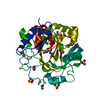
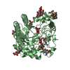
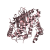
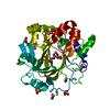
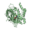
 Mycobacterium fortuitum subsp. fortuitum DSM 46621 = ATCC 6841 (bacteria)
 X-RAY DIFFRACTION /
  Authors
 United States, 3items
  Citation
 Structure visualization
 Downloads & links
 Other downloads
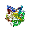


 PDBj
PDBj Assembly
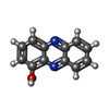
 Mass: 196.205 Da / Num. of mol.: 3 / Source method: obtained synthetically / Formula: C12H8N2O
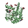
 Mass: 40.078 Da / Num. of mol.: 3 / Source method: obtained synthetically / Formula: Ca
 Mass: 18.015 Da / Num. of mol.: 213 / Source method: isolated from a natural source / Formula: H2O
 Sample preparation
 Processing