 Movie
Movie Controller

[English] 日本語
 Yorodumi
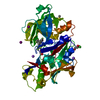
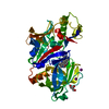
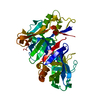
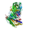
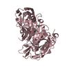
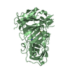
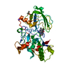
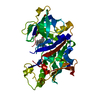
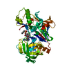
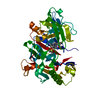
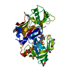
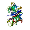
- PDB-5i3v: Crystal structure of BACE1 in complex with aminoquinoline compound 1 -
+ Open data

- Basic information
| Entry | Database: PDB / ID: 5i3v | ||||||
|---|---|---|---|---|---|---|---|
| Title | Crystal structure of BACE1 in complex with aminoquinoline compound 1 | ||||||
 Components Components | Beta-secretase 1 | ||||||
 Keywords Keywords | Hydrolase/Hydrolase Inhibitor / aspartic protease / amyloid precursor protein / Alzheimer's disease / Hydrolase-Hydrolase Inhibitor complex | ||||||
| Function / homology |  Function and homology information Function and homology informationmemapsin 2 / Golgi-associated vesicle lumen / beta-aspartyl-peptidase activity / signaling receptor ligand precursor processing / amyloid-beta formation / amyloid precursor protein catabolic process / membrane protein ectodomain proteolysis / amyloid-beta metabolic process / detection of mechanical stimulus involved in sensory perception of pain / prepulse inhibition ...memapsin 2 / Golgi-associated vesicle lumen / beta-aspartyl-peptidase activity / signaling receptor ligand precursor processing / amyloid-beta formation / amyloid precursor protein catabolic process / membrane protein ectodomain proteolysis / amyloid-beta metabolic process / detection of mechanical stimulus involved in sensory perception of pain / prepulse inhibition / cellular response to manganese ion / multivesicular body / presynaptic modulation of chemical synaptic transmission / protein serine/threonine kinase binding / cellular response to copper ion / hippocampal mossy fiber to CA3 synapse / trans-Golgi network / recycling endosome / protein processing / response to lead ion / cellular response to amyloid-beta / synaptic vesicle / late endosome / peptidase activity / positive regulation of neuron apoptotic process / amyloid-beta binding / endopeptidase activity / amyloid fibril formation / aspartic-type endopeptidase activity / early endosome / lysosome / endosome membrane / endosome / membrane raft / endoplasmic reticulum lumen / Amyloid fiber formation / axon / neuronal cell body / dendrite / enzyme binding / cell surface / Golgi apparatus / proteolysis / membrane / plasma membrane Similarity search - Function | ||||||
| Biological species |  Homo sapiens (human) Homo sapiens (human) | ||||||
| Method |  X-RAY DIFFRACTION / SYNCHROTRON / MOLECULAR REPLACEMENT / Resolution: 1.62 Å X-RAY DIFFRACTION / SYNCHROTRON / MOLECULAR REPLACEMENT / Resolution: 1.62 Å | ||||||
 Authors Authors | Whittington, D.A. / Long, A.M. | ||||||
 Citation Citation | Journal: J.Med.Chem. / Year: 2016 Title: Fragment-Linking Approach Using (19)F NMR Spectroscopy To Obtain Highly Potent and Selective Inhibitors of beta-Secretase. Authors: Jordan, J.B. / Whittington, D.A. / Bartberger, M.D. / Sickmier, E.A. / Chen, K. / Cheng, Y. / Judd, T. | ||||||
| History |
|
- Structure visualization
| Structure viewer | Molecule: MolmilJmol/JSmol |
|---|
- Downloads & links
-Download
| PDBx/mmCIF format | 5i3v.cif.gz | 102.7 KB | Display | PDBx/mmCIF format |
|---|---|---|---|---|
| PDB format | pdb5i3v.ent.gz | 74.8 KB | Display | PDB format |
| PDBx/mmJSON format | 5i3v.json.gz | Tree view | PDBx/mmJSON format | |
| Others |  Other downloads Other downloads |
-Validation report
| Summary document | 5i3v_validation.pdf.gz | 800.1 KB | Display | wwPDB validaton report |
|---|---|---|---|---|
| Full document | 5i3v_full_validation.pdf.gz | 801.8 KB | Display | |
| Data in XML | 5i3v_validation.xml.gz | 20 KB | Display | |
| Data in CIF | 5i3v_validation.cif.gz | 31.2 KB | Display | |
| Arichive directory | https://data.pdbj.org/pub/pdb/validation_reports/i3/5i3vftp://data.pdbj.org/pub/pdb/validation_reports/i3/5i3v | HTTPS FTP |
-Related structure data
| Related structure data | 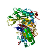 5i3wC  5i3xC 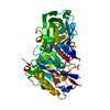 5i3yC 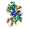 5ie1C 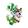 1w50S C: citing same article ( S: Starting model for refinement |
|---|---|
| Similar structure data |


-Links
 PDBj
PDBj

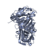
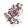
- Assembly
| Deposited unit | 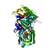
| ||||||||||||
|---|---|---|---|---|---|---|---|---|---|---|---|---|---|
| 1 |
| ||||||||||||
| Unit cell |
| ||||||||||||
| Components on special symmetry positions |
| ||||||||||||
| Details | Monomer by size exclusion chromatography |
-Components
| #1: Protein | Mass: 45822.445 Da / Num. of mol.: 1 / Fragment: UNP residues 43-453 / Mutation: R56K, R57K Source method: isolated from a genetically manipulated source Source: (gene. exp.) Homo sapiens (human) / Gene: BACE1, BACE, KIAA1149 / Production host:  | ||||||||
|---|---|---|---|---|---|---|---|---|---|
| #2: Chemical |   Mass: 126.904 Da / Num. of mol.: 3 / Source method: obtained synthetically / Formula: I Mass: 126.904 Da / Num. of mol.: 3 / Source method: obtained synthetically / Formula: I#3: Chemical | ChemComp-68M / ( | 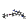  Mass: 404.548 Da / Num. of mol.: 1 / Source method: obtained synthetically / Formula: C25H32N4O Mass: 404.548 Da / Num. of mol.: 1 / Source method: obtained synthetically / Formula: C25H32N4O#4: Chemical | ChemComp-GOL / |   Mass: 92.094 Da / Num. of mol.: 1 / Source method: obtained synthetically / Formula: C3H8O3 Mass: 92.094 Da / Num. of mol.: 1 / Source method: obtained synthetically / Formula: C3H8O3#5: Water | ChemComp-HOH / |  Mass: 18.015 Da / Num. of mol.: 454 / Source method: isolated from a natural source / Formula: H2O Mass: 18.015 Da / Num. of mol.: 454 / Source method: isolated from a natural source / Formula: H2OHas protein modification | Y | |
-Experimental details
-Experiment
| Experiment | Method: X-RAY DIFFRACTION / Number of used crystals: 1 |
|---|
- Sample preparation
| Crystal | Density Matthews: 2.84 Å3/Da / Density % sol: 56.76 % |
|---|---|
| Crystal grow | Temperature: 298 K / Method: vapor diffusion / pH: 6.6 Details: 20% polyethylene glycol 5000 MME, 0.2 M ammonium iodide, 0.17 M sodium citrate (pH 6.6), 3% DMSO |
-Data collection
| Diffraction | Mean temperature: 100 K | ||||||||||||||||||||||||||||||||||||||||||||||||||||||||||||||||||
|---|---|---|---|---|---|---|---|---|---|---|---|---|---|---|---|---|---|---|---|---|---|---|---|---|---|---|---|---|---|---|---|---|---|---|---|---|---|---|---|---|---|---|---|---|---|---|---|---|---|---|---|---|---|---|---|---|---|---|---|---|---|---|---|---|---|---|---|
| Diffraction source | Source: SYNCHROTRON / Site: ALS  / Beamline: 5.0.2 / Wavelength: 1 Å / Beamline: 5.0.2 / Wavelength: 1 Å | ||||||||||||||||||||||||||||||||||||||||||||||||||||||||||||||||||
| Detector | Type: ADSC QUANTUM 315r / Detector: CCD / Date: May 18, 2012 | ||||||||||||||||||||||||||||||||||||||||||||||||||||||||||||||||||
| Radiation | Protocol: SINGLE WAVELENGTH / Monochromatic (M) / Laue (L): M / Scattering type: x-ray | ||||||||||||||||||||||||||||||||||||||||||||||||||||||||||||||||||
| Radiation wavelength | Wavelength: 1 Å / Relative weight: 1 | ||||||||||||||||||||||||||||||||||||||||||||||||||||||||||||||||||
| Reflection | Resolution: 1.62→50 Å / Num. obs: 68266 / % possible obs: 100 % / Observed criterion σ(I): -3 / Redundancy: 16.1 % / Biso Wilson estimate: 14.3 Å2 / Rmerge(I) obs: 0.067 / Net I/av σ(I): 27 / Net I/σ(I): 21.8 | ||||||||||||||||||||||||||||||||||||||||||||||||||||||||||||||||||
| Reflection shell |
|
- Processing
| Software |
| ||||||||||||||||||||||||||||||||||||||||||||||||||||||||||||
|---|---|---|---|---|---|---|---|---|---|---|---|---|---|---|---|---|---|---|---|---|---|---|---|---|---|---|---|---|---|---|---|---|---|---|---|---|---|---|---|---|---|---|---|---|---|---|---|---|---|---|---|---|---|---|---|---|---|---|---|---|---|
| Refinement | Method to determine structure: MOLECULAR REPLACEMENT Starting model: 1W50 Resolution: 1.62→50 Å / Cor.coef. Fo:Fc: 0.944 / Cor.coef. Fo:Fc free: 0.933 / SU B: 1.669 / SU ML: 0.058 / SU R Cruickshank DPI: 0.0893 / Cross valid method: THROUGHOUT / σ(F): 0 / ESU R: 0.089 / ESU R Free: 0.088 Details: HYDROGENS HAVE BEEN ADDED IN THE RIDING POSITIONS U VALUES: REFINED INDIVIDUALLY
| ||||||||||||||||||||||||||||||||||||||||||||||||||||||||||||
| Solvent computation | Ion probe radii: 0.8 Å / Shrinkage radii: 0.8 Å / VDW probe radii: 1.2 Å | ||||||||||||||||||||||||||||||||||||||||||||||||||||||||||||
| Displacement parameters | Biso max: 77.78 Å2 / Biso mean: 16.294 Å2 / Biso min: 5 Å2
| ||||||||||||||||||||||||||||||||||||||||||||||||||||||||||||
| Refinement step | Cycle: final / Resolution: 1.62→50 Å
| ||||||||||||||||||||||||||||||||||||||||||||||||||||||||||||
| Refine LS restraints |
| ||||||||||||||||||||||||||||||||||||||||||||||||||||||||||||
| LS refinement shell | Resolution: 1.62→1.662 Å / Total num. of bins used: 20
|
