 Movie
Movie Controller

[English] 日本語
 Yorodumi
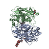
- PDB-5ewl: CRYSTAL STRUCTURE OF AMINO TERMINAL DOMAINS OF THE NMDA RECEPTOR ... -
+ Open data

- Basic information
| Entry | Database: PDB / ID: 5ewl | |||||||||
|---|---|---|---|---|---|---|---|---|---|---|
| Title | CRYSTAL STRUCTURE OF AMINO TERMINAL DOMAINS OF THE NMDA RECEPTOR SUBUNIT GLUN1 AND GLUN2B IN COMPLEX WITH MK-22 | |||||||||
 Components Components |
| |||||||||
 Keywords Keywords | TRANSPORT PROTEIN / NMDA Receptor / allosteric inhibition / GluN2B subunit | |||||||||
| Function / homology |  Function and homology information Function and homology informationexcitatory chemical synaptic transmission / Activated NTRK2 signals through FYN / Synaptic adhesion-like molecules / positive regulation of cysteine-type endopeptidase activity / negative regulation of dendritic spine maintenance / regulation of monoatomic cation transmembrane transport / glutamate receptor signaling pathway / Assembly and cell surface presentation of NMDA receptors / NMDA glutamate receptor activity / Neurexins and neuroligins ...excitatory chemical synaptic transmission / Activated NTRK2 signals through FYN / Synaptic adhesion-like molecules / positive regulation of cysteine-type endopeptidase activity / negative regulation of dendritic spine maintenance / regulation of monoatomic cation transmembrane transport / glutamate receptor signaling pathway / Assembly and cell surface presentation of NMDA receptors / NMDA glutamate receptor activity / Neurexins and neuroligins / NMDA selective glutamate receptor complex / glutamate-gated calcium ion channel activity / calcium ion transmembrane import into cytosol / protein heterotetramerization / glutamate binding / glycine binding / response to zinc ion / Negative regulation of NMDA receptor-mediated neuronal transmission / Unblocking of NMDA receptors, glutamate binding and activation / monoatomic cation transmembrane transport / regulation of neuronal synaptic plasticity / positive regulation of excitatory postsynaptic potential / Long-term potentiation / MECP2 regulates neuronal receptors and channels / ligand-gated monoatomic ion channel activity involved in regulation of presynaptic membrane potential / EPHB-mediated forward signaling / ionotropic glutamate receptor signaling pathway / Ras activation upon Ca2+ influx through NMDA receptor / excitatory postsynaptic potential / positive regulation of synaptic transmission, glutamatergic / synaptic membrane / synaptic transmission, glutamatergic / transmitter-gated monoatomic ion channel activity involved in regulation of postsynaptic membrane potential / regulation of membrane potential / long-term synaptic potentiation / postsynaptic density membrane / regulation of synaptic plasticity / brain development / late endosome / amyloid-beta binding / RAF/MAP kinase cascade / chemical synaptic transmission / postsynaptic membrane / response to ethanol / postsynaptic density / learning or memory / lysosome / cytoskeleton / neuron projection / endoplasmic reticulum membrane / cell surface / zinc ion binding / metal ion binding / plasma membrane / cytoplasm Similarity search - Function | |||||||||
| Biological species |  Homo sapiens (human) Homo sapiens (human) | |||||||||
| Method |  X-RAY DIFFRACTION / SYNCHROTRON / FOURIER SYNTHESIS / Resolution: 2.98 Å X-RAY DIFFRACTION / SYNCHROTRON / FOURIER SYNTHESIS / Resolution: 2.98 Å | |||||||||
 Authors Authors | Pandit, J. | |||||||||
 Citation Citation | Journal: Mol.Pharmacol. / Year: 2016 Title: A Novel Binding Mode Reveals Two Distinct Classes of NMDA Receptor GluN2B-selective Antagonists. Authors: Stroebel, D. / Buhl, D.L. / Knafels, J.D. / Chanda, P.K. / Green, M. / Sciabola, S. / Mony, L. / Paoletti, P. / Pandit, J. | |||||||||
| History |
|
- Structure visualization
| Structure viewer | Molecule: MolmilJmol/JSmol |
|---|
- Downloads & links
-Download
| PDBx/mmCIF format | 5ewl.cif.gz | 580.7 KB | Display | PDBx/mmCIF format |
|---|---|---|---|---|
| PDB format | pdb5ewl.ent.gz | 476.9 KB | Display | PDB format |
| PDBx/mmJSON format | 5ewl.json.gz | Tree view | PDBx/mmJSON format | |
| Others |  Other downloads Other downloads |
-Validation report
| Summary document | 5ewl_validation.pdf.gz | 1.2 MB | Display | wwPDB validaton report |
|---|---|---|---|---|
| Full document | 5ewl_full_validation.pdf.gz | 1.2 MB | Display | |
| Data in XML | 5ewl_validation.xml.gz | 53.1 KB | Display | |
| Data in CIF | 5ewl_validation.cif.gz | 73.3 KB | Display | |
| Arichive directory | https://data.pdbj.org/pub/pdb/validation_reports/ew/5ewlftp://data.pdbj.org/pub/pdb/validation_reports/ew/5ewl | HTTPS FTP |
-Related structure data
| Related structure data |  5ewjC 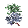 5ewmC 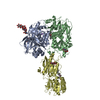 3qelS C: citing same article ( S: Starting model for refinement |
|---|---|
| Similar structure data |


-Links
 PDBj
PDBj


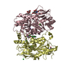
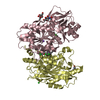
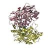
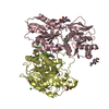
- Assembly
| Deposited unit | 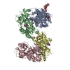
| ||||||||
|---|---|---|---|---|---|---|---|---|---|
| 1 | 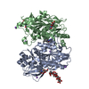
| ||||||||
| 2 | 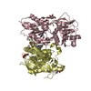
| ||||||||
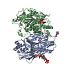
| Unit cell |
|
-Components
-Protein , 2 types, 4 molecules ACBD
| #1: Protein | Mass: 43757.977 Da / Num. of mol.: 2 / Fragment: Amino Terminal Domain (UNP residues 23-408) / Mutation: N61Q,N371Q Source method: isolated from a genetically manipulated source Source: (gene. exp.)   Spodoptera frugiperda (fall armyworm) / References: UniProt: Q91977, UniProt: A0A1L8F5J9*PLUS Spodoptera frugiperda (fall armyworm) / References: UniProt: Q91977, UniProt: A0A1L8F5J9*PLUS#2: Protein | Mass: 41351.902 Da / Num. of mol.: 2 / Fragment: Amino Terminal Domain (UNP residues 31-394) / Mutation: N348D Source method: isolated from a genetically manipulated source Source: (gene. exp.) Homo sapiens (human) / Gene: GRIN2B, NMDAR2B / Production host: Spodoptera frugiperda (fall armyworm) / References: UniProt: Q13224 |
|---|
-Sugars , 2 types, 7 molecules 
| #3: Polysaccharide | alpha-D-mannopyranose-(1-3)-[alpha-D-mannopyranose-(1-6)]beta-D-mannopyranose-(1-4)-2-acetamido-2- ...alpha-D-mannopyranose-(1-3)-[alpha-D-mannopyranose-(1-6)]beta-D-mannopyranose-(1-4)-2-acetamido-2-deoxy-beta-D-glucopyranose-(1-4)-2-acetamido-2-deoxy-beta-D-glucopyranose Source method: isolated from a genetically manipulated source |
|---|---|
| #5: Sugar | ChemComp-NAG /  Type: D-saccharide, beta linking / Mass: 221.208 Da / Num. of mol.: 6 Type: D-saccharide, beta linking / Mass: 221.208 Da / Num. of mol.: 6Source method: isolated from a genetically manipulated source Formula: C8H15NO6 |
-Non-polymers , 3 types, 277 molecules 
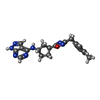

| #4: Chemical | ChemComp-NA /  Mass: 22.990 Da / Num. of mol.: 6 / Source method: obtained synthetically / Formula: Na Mass: 22.990 Da / Num. of mol.: 6 / Source method: obtained synthetically / Formula: Na#6: Chemical |  Mass: 375.427 Da / Num. of mol.: 2 / Source method: obtained synthetically / Formula: C20H21N7O Mass: 375.427 Da / Num. of mol.: 2 / Source method: obtained synthetically / Formula: C20H21N7O#7: Water | ChemComp-HOH / | Mass: 18.015 Da / Num. of mol.: 269 / Source method: isolated from a natural source / Formula: H2O |
|---|
-Experimental details
-Experiment
| Experiment | Method: X-RAY DIFFRACTION / Number of used crystals: 1 |
|---|
- Sample preparation
| Crystal | Density Matthews: 3.06 Å3/Da / Density % sol: 59.79 % |
|---|---|
| Crystal grow | Temperature: 298 K / Method: vapor diffusion, hanging drop / Details: 3.6M Na Formate, 0.1 M Hepes pH 7.0 / PH range: 6.8-7.2 |
-Data collection
| Diffraction | Mean temperature: 100 K | |||||||||||||||||||||||||||
|---|---|---|---|---|---|---|---|---|---|---|---|---|---|---|---|---|---|---|---|---|---|---|---|---|---|---|---|---|
| Diffraction source | Source: SYNCHROTRON / Site: APS  / Beamline: 17-ID / Wavelength: 1 Å / Beamline: 17-ID / Wavelength: 1 Å | |||||||||||||||||||||||||||
| Detector | Type: DECTRIS PILATUS3 6M / Detector: PIXEL / Date: Aug 20, 2014 | |||||||||||||||||||||||||||
| Radiation | Protocol: SINGLE WAVELENGTH / Monochromatic (M) / Laue (L): M / Scattering type: x-ray | |||||||||||||||||||||||||||
| Radiation wavelength | Wavelength: 1 Å / Relative weight: 1 | |||||||||||||||||||||||||||
| Reflection | Resolution: 2.98→120.31 Å / Num. obs: 41141 / % possible obs: 96.2 % / Redundancy: 2.6 % / Biso Wilson estimate: 80.34 Å2 / CC1/2: 0.998 / Rmerge(I) obs: 0.054 / Rpim(I) all: 0.038 / Net I/σ(I): 14.3 / Num. measured all: 106198 | |||||||||||||||||||||||||||
| Reflection shell | Diffraction-ID: 1 / Rejects: _
|
- Processing
| Software |
| |||||||||||||||||||||||||||||||||||||||||||||||||||||||||||||||||||||||||||||||||||||||||||||||||||||||||||||||||||||||||||||
|---|---|---|---|---|---|---|---|---|---|---|---|---|---|---|---|---|---|---|---|---|---|---|---|---|---|---|---|---|---|---|---|---|---|---|---|---|---|---|---|---|---|---|---|---|---|---|---|---|---|---|---|---|---|---|---|---|---|---|---|---|---|---|---|---|---|---|---|---|---|---|---|---|---|---|---|---|---|---|---|---|---|---|---|---|---|---|---|---|---|---|---|---|---|---|---|---|---|---|---|---|---|---|---|---|---|---|---|---|---|---|---|---|---|---|---|---|---|---|---|---|---|---|---|---|---|---|
| Refinement | Method to determine structure: FOURIER SYNTHESIS Starting model: 3QEL Resolution: 2.98→30.04 Å / Cor.coef. Fo:Fc: 0.9424 / Cor.coef. Fo:Fc free: 0.9 / Cross valid method: THROUGHOUT / σ(F): 0 / SU Rfree Blow DPI: 0.339
| |||||||||||||||||||||||||||||||||||||||||||||||||||||||||||||||||||||||||||||||||||||||||||||||||||||||||||||||||||||||||||||
| Displacement parameters | Biso mean: 72.87 Å2
| |||||||||||||||||||||||||||||||||||||||||||||||||||||||||||||||||||||||||||||||||||||||||||||||||||||||||||||||||||||||||||||
| Refine analyze | Luzzati coordinate error obs: 0.308 Å | |||||||||||||||||||||||||||||||||||||||||||||||||||||||||||||||||||||||||||||||||||||||||||||||||||||||||||||||||||||||||||||
| Refinement step | Cycle: LAST / Resolution: 2.98→30.04 Å
| |||||||||||||||||||||||||||||||||||||||||||||||||||||||||||||||||||||||||||||||||||||||||||||||||||||||||||||||||||||||||||||
| Refine LS restraints |
| |||||||||||||||||||||||||||||||||||||||||||||||||||||||||||||||||||||||||||||||||||||||||||||||||||||||||||||||||||||||||||||
| LS refinement shell | Resolution: 2.98→3.06 Å / Total num. of bins used: 20
| |||||||||||||||||||||||||||||||||||||||||||||||||||||||||||||||||||||||||||||||||||||||||||||||||||||||||||||||||||||||||||||
| Refinement TLS params. | Method: refined / Refine-ID: X-RAY DIFFRACTION
| |||||||||||||||||||||||||||||||||||||||||||||||||||||||||||||||||||||||||||||||||||||||||||||||||||||||||||||||||||||||||||||
| Refinement TLS group |
|
