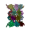
Movie
Movie Controller

[English] 日本語
 Yorodumi
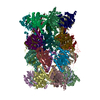
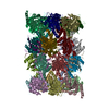
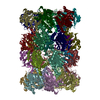
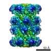
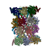
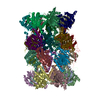
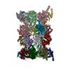
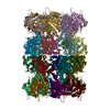
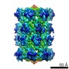
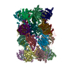
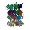
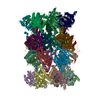
- PDB-5a0q: Cryo-EM reveals the conformation of a substrate analogue in the h... -
+ Open data

- Basic information
| Entry | Database: PDB / ID: 5a0q | ||||||
|---|---|---|---|---|---|---|---|
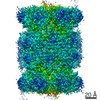
| Title | Cryo-EM reveals the conformation of a substrate analogue in the human 20S proteasome core | ||||||
 Components Components |
| ||||||
 Keywords Keywords | HYDROLASE / PROTEASOME / 20S / ADAAHX3L3VS / LIGAND / INHIBITOR / DRUG DESIGN | ||||||
| Function / homology |  Function and homology information Function and homology informationpurine ribonucleoside triphosphate binding / CD8-positive, alpha-beta T cell differentiation / CD8-positive, alpha-beta T cell homeostasis / thymic T cell selection / Antigen processing: Ub, ATP-independent proteasomal degradation / structural constituent of proteasome / spermatoproteasome complex / negative regulation of regulatory T cell differentiation / T-helper 1 cell differentiation / Regulation of ornithine decarboxylase (ODC) ...purine ribonucleoside triphosphate binding / CD8-positive, alpha-beta T cell differentiation / CD8-positive, alpha-beta T cell homeostasis / thymic T cell selection / Antigen processing: Ub, ATP-independent proteasomal degradation / structural constituent of proteasome / spermatoproteasome complex / negative regulation of regulatory T cell differentiation / T-helper 1 cell differentiation / Regulation of ornithine decarboxylase (ODC) / proteasome core complex / Proteasome assembly / cellular response to type I interferon / T-helper 17 cell differentiation / Cross-presentation of soluble exogenous antigens (endosomes) / Somitogenesis / flagellated sperm motility / sperm end piece / immune system process / myofibril / proteasomal ubiquitin-independent protein catabolic process / ciliary tip / proteasome endopeptidase complex / NF-kappaB binding / proteasome core complex, beta-subunit complex / threonine-type endopeptidase activity / proteasome core complex, alpha-subunit complex / proteasome assembly / regulation of G1/S transition of mitotic cell cycle / positive regulation of interleukin-2 production / proteasome storage granule / response to type II interferon / proteasome complex / regulation of proteasomal protein catabolic process / meiotic cell cycle / sarcomere / Regulation of activated PAK-2p34 by proteasome mediated degradation / negative regulation of inflammatory response to antigenic stimulus / proteasomal protein catabolic process / Autodegradation of Cdh1 by Cdh1:APC/C / APC/C:Cdc20 mediated degradation of Securin / lipopolysaccharide binding / Asymmetric localization of PCP proteins / Ubiquitin-dependent degradation of Cyclin D / positive regulation of type II interferon production / SCF-beta-TrCP mediated degradation of Emi1 / AUF1 (hnRNP D0) binds and destabilizes mRNA / NIK-->noncanonical NF-kB signaling / P-body / TNFR2 non-canonical NF-kB pathway / protein catabolic process / Assembly of the pre-replicative complex / Vpu mediated degradation of CD4 / Cdc20:Phospho-APC/C mediated degradation of Cyclin A / Dectin-1 mediated noncanonical NF-kB signaling / Degradation of DVL / Degradation of AXIN / Degradation of CRY and PER proteins / Hh mutants are degraded by ERAD / Activation of NF-kappaB in B cells / G2/M Checkpoints / Degradation of GLI1 by the proteasome / Hedgehog ligand biogenesis / Regulation of RUNX3 expression and activity / Autodegradation of the E3 ubiquitin ligase COP1 / Defective CFTR causes cystic fibrosis / GSK3B and BTRC:CUL1-mediated-degradation of NFE2L2 / response to virus / Negative regulation of NOTCH4 signaling / AMPK-induced ERAD and lysosome mediated degradation of PD-L1(CD274) / Hedgehog 'on' state / APC/C:Cdh1 mediated degradation of Cdc20 and other APC/C:Cdh1 targeted proteins in late mitosis/early G1 / Vif-mediated degradation of APOBEC3G / FBXL7 down-regulates AURKA during mitotic entry and in early mitosis / Degradation of GLI2 by the proteasome / GLI3 is processed to GLI3R by the proteasome / MAPK6/MAPK4 signaling / GSK3B-mediated proteasomal degradation of PD-L1(CD274) / Degradation of CDH1 / Degradation of beta-catenin by the destruction complex / Oxygen-dependent proline hydroxylation of Hypoxia-inducible Factor Alpha / CDK-mediated phosphorylation and removal of Cdc6 / ABC-family protein mediated transport / CLEC7A (Dectin-1) signaling / Activation of STAT3 by cadherin engagement / SCF(Skp2)-mediated degradation of p27/p21 / FCERI mediated NF-kB activation / nuclear matrix / SPOP-mediated proteasomal degradation of PD-L1(CD274) / Regulation of expression of SLITs and ROBOs / Regulation of PTEN stability and activity / Ubiquitin-Mediated Degradation of Phosphorylated Cdc25A / Ribosome Quality Control (RQC) complex extracts and degrades nascent peptide / Interleukin-1 signaling / Orc1 removal from chromatin / peptidase activity / positive regulation of tumor necrosis factor production / Regulation of RUNX2 expression and activity / Regulation of RAS by GAPs / The role of GTSE1 in G2/M progression after G2 checkpoint Similarity search - Function | ||||||
| Biological species |  HOMO SAPIENS (human) HOMO SAPIENS (human) | ||||||
| Method | ELECTRON MICROSCOPY / single particle reconstruction / cryo EM / Resolution: 3.5 Å | ||||||
 Authors Authors | daFonseca, P.C.A. / Morris, E.P. | ||||||
 Citation Citation | Journal: Nat Commun / Year: 2015 Title: Cryo-EM reveals the conformation of a substrate analogue in the human 20S proteasome core. Authors: Paula C A da Fonseca / Edward P Morris /  Abstract: The proteasome is a highly regulated protease complex fundamental for cell homeostasis and controlled cell cycle progression. It functions by removing a wide range of specifically tagged proteins, ...The proteasome is a highly regulated protease complex fundamental for cell homeostasis and controlled cell cycle progression. It functions by removing a wide range of specifically tagged proteins, including key cellular regulators. Here we present the structure of the human 20S proteasome core bound to a substrate analogue inhibitor molecule, determined by electron cryo-microscopy (cryo-EM) and single-particle analysis at a resolution of around 3.5 Å. Our map allows the building of protein coordinates as well as defining the location and conformation of the inhibitor at the different active sites. These results open new prospects to tackle the proteasome functional mechanisms. Moreover, they also further demonstrate that cryo-EM is emerging as a realistic approach for general structural studies of protein-ligand interactions. | ||||||
| History |
|
- Structure visualization
| Movie |
Movie viewer |
|---|---|
| Structure viewer | Molecule: MolmilJmol/JSmol |
- Downloads & links
-Download
| PDBx/mmCIF format | 5a0q.cif.gz | 943.4 KB | Display | PDBx/mmCIF format |
|---|---|---|---|---|
| PDB format | pdb5a0q.ent.gz | 739.7 KB | Display | PDB format |
| PDBx/mmJSON format | 5a0q.json.gz | Tree view | PDBx/mmJSON format | |
| Others |  Other downloads Other downloads |
-Validation report
| Arichive directory | https://data.pdbj.org/pub/pdb/validation_reports/a0/5a0qftp://data.pdbj.org/pub/pdb/validation_reports/a0/5a0q | HTTPS FTP |
|---|
-Related structure data
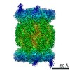
| Related structure data | 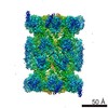 2981MC M: map data used to model this data C: citing same article ( |
|---|---|
| Similar structure data | |
| EM raw data | EMPIAR-10038 (Title: Cryo-EM reveals the conformation of a substrate analogue in the human 20S proteasome core Data size: 579.1 Data #1: raw micrographs of the human 20S proteasome core complex bound to the ligand AdaAhx3L3VS [micrographs - multiframe]) |
-Links
 PDBj
PDBj



- Assembly
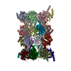
| Deposited unit | 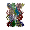
|
|---|---|
| 1 |
|
-Components
-PROTEASOME SUBUNIT ALPHA TYPE- ... , 7 types, 14 molecules AOBPCQDRESFTGU
| #1: Protein | Mass: 27432.459 Da / Num. of mol.: 2 / Source method: isolated from a natural source / Source: (natural) HOMO SAPIENS (human) / Cell: ERYTHROCYTESReferences: UniProt: P60900, proteasome endopeptidase complex #2: Protein | Mass: 25927.535 Da / Num. of mol.: 2 / Source method: isolated from a natural source / Source: (natural) HOMO SAPIENS (human) / Cell: ERYTHROCYTESReferences: UniProt: P25787, proteasome endopeptidase complex #3: Protein | Mass: 29525.842 Da / Num. of mol.: 2 / Source method: isolated from a natural source / Source: (natural) HOMO SAPIENS (human) / Cell: ERYTHROCYTESReferences: UniProt: P25789, proteasome endopeptidase complex #4: Protein | Mass: 27929.891 Da / Num. of mol.: 2 / Source method: isolated from a natural source / Source: (natural) HOMO SAPIENS (human) / Cell: ERYTHROCYTESReferences: UniProt: O14818, proteasome endopeptidase complex #5: Protein | Mass: 26435.977 Da / Num. of mol.: 2 / Source method: isolated from a natural source / Source: (natural) HOMO SAPIENS (human) / Cell: ERYTHROCYTESReferences: UniProt: P28066, proteasome endopeptidase complex #6: Protein | Mass: 29595.627 Da / Num. of mol.: 2 / Source method: isolated from a natural source / Source: (natural) HOMO SAPIENS (human) / Cell: ERYTHROCYTESReferences: UniProt: P25786, proteasome endopeptidase complex #7: Protein | Mass: 28469.252 Da / Num. of mol.: 2 / Source method: isolated from a natural source / Source: (natural) HOMO SAPIENS (human) / Cell: ERYTHROCYTESReferences: UniProt: P25788, proteasome endopeptidase complex |
|---|
-PROTEASOME SUBUNIT BETA TYPE- ... , 7 types, 14 molecules HVIWJXKYLZMaNb
| #8: Protein | Mass: 21921.836 Da / Num. of mol.: 2 / Source method: isolated from a natural source Details: SUBUNIT PARTIALLY BOUND TO THE INHIBITOR ADAMANTANE-ACETYL-(6-AMINOHEXANOYL)3-(LEUCINYL)3-VINYL-(METHYL)-SULFONE (ADAAHX3L3VS) Source: (natural) HOMO SAPIENS (human) / Cell: ERYTHROCYTESReferences: UniProt: P28072, proteasome endopeptidase complex #9: Protein | Mass: 25321.980 Da / Num. of mol.: 2 / Source method: isolated from a natural source Details: SUBUNIT PARTIALLY BOUND TO THE INHIBITOR ADAMANTANE-ACETYL-(6-AMINOHEXANOYL)3-(LEUCINYL)3-VINYL-(METHYL)-SULFONE (ADAAHX3L3VS) Source: (natural) HOMO SAPIENS (human) / Cell: ERYTHROCYTESReferences: UniProt: Q99436, proteasome endopeptidase complex #10: Protein | Mass: 22841.701 Da / Num. of mol.: 2 / Source method: isolated from a natural source / Source: (natural) HOMO SAPIENS (human) / Cell: ERYTHROCYTESReferences: UniProt: P49720, proteasome endopeptidase complex #11: Protein | Mass: 22864.277 Da / Num. of mol.: 2 / Source method: isolated from a natural source / Source: (natural) HOMO SAPIENS (human) / Cell: ERYTHROCYTESReferences: UniProt: P49721, proteasome endopeptidase complex #12: Protein | Mass: 22484.369 Da / Num. of mol.: 2 / Source method: isolated from a natural source Details: SUBUNIT BOUND TO THE INHIBITOR ADAMANTANE-ACETYL-(6-AMINOHEXANOYL)3-(LEUCINYL)3-VINYL-(METHYL)-SULFONE (ADAAHX3L3VS) Source: (natural) HOMO SAPIENS (human) / Cell: ERYTHROCYTESReferences: UniProt: P28074, proteasome endopeptidase complex #13: Protein | Mass: 23578.986 Da / Num. of mol.: 2 / Source method: isolated from a natural source / Source: (natural) HOMO SAPIENS (human) / Cell: ERYTHROCYTESReferences: UniProt: P20618, proteasome endopeptidase complex #14: Protein | Mass: 24414.740 Da / Num. of mol.: 2 / Source method: isolated from a natural source / Source: (natural) HOMO SAPIENS (human) / Cell: ERYTHROCYTESReferences: UniProt: P28070, proteasome endopeptidase complex |
|---|
-Non-polymers , 1 types, 6 molecules 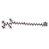
| #15: Chemical | ChemComp-KNM /  Mass: 949.377 Da / Num. of mol.: 6 / Source method: obtained synthetically / Formula: C51H92N6O8S Mass: 949.377 Da / Num. of mol.: 6 / Source method: obtained synthetically / Formula: C51H92N6O8S |
|---|
-Details
| Has protein modification | Y |
|---|
-Experimental details
-Experiment
| Experiment | Method: ELECTRON MICROSCOPY |
|---|---|
| EM experiment | Aggregation state: PARTICLE / 3D reconstruction method: single particle reconstruction |
- Sample preparation
| Component | Name: HUMAN 20S PROTEASOME CORE / Type: COMPLEX Details: THE POWER SPECTRA OF THE IMAGES SELECTED FOR ANALYSIS HAD ISOTROPIC THON RINGS DIRECTLY OBSERVED TO 4 ANGSTROMS OR BETTER |
|---|---|
| Buffer solution | Name: 50 MM TRIS-HCL, 5 MM MGCL2 AND 1MM DITHIOTREITOL / pH: 7.5 / Details: 50 MM TRIS-HCL, 5 MM MGCL2 AND 1MM DITHIOTREITOL |
| Specimen | Conc.: 0.1 mg/ml / Embedding applied: NO / Shadowing applied: NO / Staining applied: NO / Vitrification applied: YES |
| Specimen support | Details: CARBON |
| Vitrification | Instrument: FEI VITROBOT MARK III / Cryogen name: ETHANE Details: VITRIFICATION 1 -- CRYOGEN- ETHANE, HUMIDITY- 95, TEMPERATURE- 120, INSTRUMENT- FEI VITROBOT MARK III, METHOD- BLOT 2. 5 SECONDS BEFORE PLUNGING, |
- Electron microscopy imaging
| Experimental equipment |  Model: Titan Krios / Image courtesy: FEI Company |
|---|---|
| Microscopy | Model: FEI TITAN KRIOS / Date: Feb 4, 2014 Details: EACH EXPOSURE WAS RECORDED AS 17 INDIVIDUAL FRAMES CAPTURED AT A RATE OF 0.056 SECONDS PER FRAME, WITH AN ELECTRON DOSE OF 2.8 ELECTRONS PER SQUARE ANGSTROM. DATA-SET RECORDED USING EPU SOFTWARE. |
| Electron gun | Electron source:  FIELD EMISSION GUN / Accelerating voltage: 300 kV / Illumination mode: FLOOD BEAM FIELD EMISSION GUN / Accelerating voltage: 300 kV / Illumination mode: FLOOD BEAM |
| Electron lens | Mode: BRIGHT FIELD / Nominal magnification: 75000 X / Calibrated magnification: 134461 X / Nominal defocus max: 3000 nm / Nominal defocus min: 1700 nm / Cs: 2.7 mm |
| Specimen holder | Temperature: 85 K |
| Image recording | Electron dose: 4.8 e/Å2 / Film or detector model: FEI FALCON II (4k x 4k) |
| Image scans | Num. digital images: 960 |
- Processing
| EM software |
| ||||||||||||||||||
|---|---|---|---|---|---|---|---|---|---|---|---|---|---|---|---|---|---|---|---|
| CTF correction | Details: FULL RECORDED IMAGE | ||||||||||||||||||
| Symmetry | Point symmetry: C2 (2 fold cyclic) | ||||||||||||||||||
| 3D reconstruction | Method: PROJECTION MATCHING / Resolution: 3.5 Å / Num. of particles: 76500 / Actual pixel size: 1.04 Å Details: DISORDERED REGIONS, NOT RECOVERED IN THE EM MAP, WHERE NOT MODELED. SUBMISSION BASED ON EXPERIMENTAL DATA FROM EMDB EMD-2981. (DEPOSITION ID: 13310). Symmetry type: POINT | ||||||||||||||||||
| Atomic model building | Protocol: FLEXIBLE FIT / Space: REAL / Details: METHOD--FLEXIBLE | ||||||||||||||||||
| Atomic model building | PDB-ID: 3UNE Accession code: 3UNE / Source name: PDB / Type: experimental model | ||||||||||||||||||
| Refinement | Highest resolution: 3.5 Å | ||||||||||||||||||
| Refinement step | Cycle: LAST / Highest resolution: 3.5 Å
|