 Movie
Movie Controller

[English] 日本語
 Yorodumi
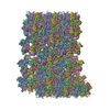
- EMDB-5136: CryoEM Structure of Tubular Assembly of HIV-1 CA and Evidence for... -
+ Open data

- Basic information
| Entry | Database: EMDB / ID: EMD-5136 | |||||||||
|---|---|---|---|---|---|---|---|---|---|---|
| Title | CryoEM Structure of Tubular Assembly of HIV-1 CA and Evidence for a Novel Interface | |||||||||
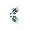
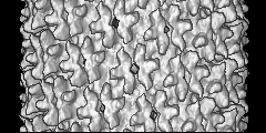
 Map data Map data | This is a map of tubular assembly of HIV-1 CA | |||||||||
 Sample Sample |
| |||||||||
| Biological species |   Human immunodeficiency virus Human immunodeficiency virus | |||||||||
| Method | helical reconstruction / cryo EM / Resolution: 16.0 Å | |||||||||
 Authors Authors | Byeon IL / Meng X / Jung J / Zhao G / Ahn J / Concel J / Aiken C / Gronenborn AM / Zhang P | |||||||||
 Citation Citation | Journal: Cell / Year: 2009 Title: Structural convergence between Cryo-EM and NMR reveals intersubunit interactions critical for HIV-1 capsid function. Authors: In-Ja L Byeon / Xin Meng / Jinwon Jung / Gongpu Zhao / Ruifeng Yang / Jinwoo Ahn / Jiong Shi / Jason Concel / Christopher Aiken / Peijun Zhang / Angela M Gronenborn /  Abstract: Mature HIV-1 particles contain conical-shaped capsids that enclose the viral RNA genome and perform essential functions in the virus life cycle. Previous structural analysis of two- and three- ...Mature HIV-1 particles contain conical-shaped capsids that enclose the viral RNA genome and perform essential functions in the virus life cycle. Previous structural analysis of two- and three-dimensional arrays of the capsid protein (CA) hexamer revealed three interfaces. Here, we present a cryoEM study of a tubular assembly of CA and a high-resolution NMR structure of the CA C-terminal domain (CTD) dimer. In the solution dimer structure, the monomers exhibit different relative orientations compared to previous X-ray structures. The solution structure fits well into the EM density map, suggesting that the dimer interface is retained in the assembled CA. We also identified a CTD-CTD interface at the local three-fold axis in the cryoEM map and confirmed its functional importance by mutagenesis. In the tubular assembly, CA intermolecular interfaces vary slightly, accommodating the asymmetry present in tubes. This provides the necessary plasticity to allow for controlled virus capsid dis/assembly. | |||||||||
| History |
|
- Structure visualization
| Movie |
 Movie viewer Movie viewer |
|---|---|
| Structure viewer | EM map: SurfViewMolmilJmol/JSmol |
| Supplemental images |

- Downloads & links
-EMDB archive
| Map data | emd_5136.map.gz | 11.1 MB | EMDB map data format | |
|---|---|---|---|---|
| Header (meta data) | emd-5136-v30.xmlemd-5136.xml | 7.4 KB 7.4 KB | Display Display | EMDB header |
| Images | emd_5136_1.tif | 732.6 KB | ||
| Archive directory |  http://ftp.pdbj.org/pub/emdb/structures/EMD-5136ftp://ftp.pdbj.org/pub/emdb/structures/EMD-5136 http://ftp.pdbj.org/pub/emdb/structures/EMD-5136ftp://ftp.pdbj.org/pub/emdb/structures/EMD-5136 | HTTPS FTP |
-Validation report
| Summary document | emd_5136_validation.pdf.gz | 78 KB | Display | EMDB validaton report |
|---|---|---|---|---|
| Full document | emd_5136_full_validation.pdf.gz | 77.1 KB | Display | |
| Data in XML | emd_5136_validation.xml.gz | 493 B | Display | |
| Arichive directory | https://ftp.pdbj.org/pub/emdb/validation_reports/EMD-5136ftp://ftp.pdbj.org/pub/emdb/validation_reports/EMD-5136 | HTTPS FTP |
-Related structure data

-Links
| EMDB pages | EMDB (EBI/PDBe) / EMDataResource |
|---|
-Map
| File | Download / File: emd_5136.map.gz / Format: CCP4 / Size: 25.8 MB / Type: IMAGE STORED AS FLOATING POINT NUMBER (4 BYTES) | ||||||||||||||||||||||||||||||||||||||||||||||||||||||||||||||||||||
|---|---|---|---|---|---|---|---|---|---|---|---|---|---|---|---|---|---|---|---|---|---|---|---|---|---|---|---|---|---|---|---|---|---|---|---|---|---|---|---|---|---|---|---|---|---|---|---|---|---|---|---|---|---|---|---|---|---|---|---|---|---|---|---|---|---|---|---|---|---|
| Annotation | This is a map of tubular assembly of HIV-1 CA | ||||||||||||||||||||||||||||||||||||||||||||||||||||||||||||||||||||
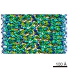
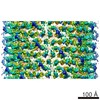
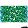
| Projections & slices | Image control
Images are generated by Spider. generated in cubic-lattice coordinate | ||||||||||||||||||||||||||||||||||||||||||||||||||||||||||||||||||||
| Voxel size | X=Y=Z: 2.15 Å | ||||||||||||||||||||||||||||||||||||||||||||||||||||||||||||||||||||
| Density |
| ||||||||||||||||||||||||||||||||||||||||||||||||||||||||||||||||||||
| Symmetry | Space group: 1 | ||||||||||||||||||||||||||||||||||||||||||||||||||||||||||||||||||||
| Details | EMDB XML:
CCP4 map header:
| ||||||||||||||||||||||||||||||||||||||||||||||||||||||||||||||||||||
 Z (Sec.)
 Y (Row.)
 X (Col.)


-Supplemental data
- Sample components
-Entire : HIV-1 CA A92E
| Entire | Name: HIV-1 CA A92E |
|---|---|
| Components |
|
-Supramolecule #1000: HIV-1 CA A92E
| Supramolecule | Name: HIV-1 CA A92E / type: sample / ID: 1000 / Details: component name, HIV-1 CA protein / Number unique components: 6 |
|---|
-Macromolecule #1: HIV-1 CA protein
| Macromolecule | Name: HIV-1 CA protein / type: protein_or_peptide / ID: 1 / Name.synonym: hexamer / Recombinant expression: Yes / Database: NCBI |
|---|---|
| Source (natural) | Organism: Human immunodeficiency virus / synonym: Human immunodeficiency virus |
-Experimental details
-Structure determination
| Method | cryo EM |
|---|---|
 Processing Processing | helical reconstruction |
| Aggregation state | filament |
-Sample preparation
| Vitrification | Cryogen name: NITROGEN / Instrument: OTHER |
|---|
- Electron microscopy
| Microscope | FEI POLARA 300 |
|---|---|
| Electron beam | Acceleration voltage: 200 kV / Electron source:  FIELD EMISSION GUN FIELD EMISSION GUN |
| Electron optics | Illumination mode: SPOT SCAN / Imaging mode: BRIGHT FIELD |
| Sample stage | Specimen holder: liquid nitrogen-cooled cryo specimen holder Specimen holder model: GATAN LIQUID NITROGEN |
| Experimental equipment |  Model: Tecnai Polara / Image courtesy: FEI Company |
-Image processing
| Final reconstruction | Resolution.type: BY AUTHOR / Resolution: 16.0 Å / Resolution method: FSC 0.5 CUT-OFF |
|---|
-Atomic model buiding 1
| Initial model | PDB ID: |
|---|---|
| Software | Name: Chimera |
| Refinement | Space: REAL |

