 ムービー
ムービー コントローラー


+ データを開く

- 基本情報
基本情報
| 登録情報 | データベース: EMDB / ID: EMD-5130 | |||||||||
|---|---|---|---|---|---|---|---|---|---|---|
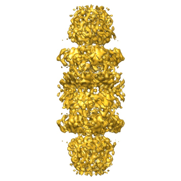
| タイトル | 3D cryo-EM Structure of Archaeal 20S Proteasome in Complex with the C-terminus of PAN | |||||||||
 マップデータ マップデータ | This is the 3D volume of T.acidophilum 20S proteasome in complex with a hybrid activator of PA26 and PAN | |||||||||
 試料 試料 |
| |||||||||
 キーワード キーワード | Proteasome / proteasomal ATPase / protein degradation / AAA ATPase / x-ray crystallography / electron cryomicroscopy | |||||||||
| 生物種 |   Thermoplasma acidophilum (好酸性) Thermoplasma acidophilum (好酸性) | |||||||||
| 手法 | 単粒子再構成法 / クライオ電子顕微鏡法 / 解像度: 7.5 Å | |||||||||
 データ登録者 データ登録者 | Yu Y / Smith DM / Kim H / Rodriguez V / Goldberg AL / Cheng Y | |||||||||
 引用 引用 | ジャーナル: EMBO J / 年: 2010 タイトル: Interactions of PAN's C-termini with archaeal 20S proteasome and implications for the eukaryotic proteasome-ATPase interactions. 著者: Yadong Yu / David M Smith / Ho Min Kim / Victor Rodriguez / Alfred L Goldberg / Yifan Cheng /  要旨: Protein degradation in the 20S proteasome is regulated in eukaryotes by the 19S ATPase complex and in archaea by the homologous PAN ATPase ring complex. Subunits of these hexameric ATPases contain on ...Protein degradation in the 20S proteasome is regulated in eukaryotes by the 19S ATPase complex and in archaea by the homologous PAN ATPase ring complex. Subunits of these hexameric ATPases contain on their C-termini a conserved hydrophobic-tyrosine-X (HbYX) motif that docks into pockets in the 20S to stimulate the opening of a gated substrate entry channel. Here, we report the crystal structure of the archaeal 20S proteasome in complex with the C-terminus of the archaeal proteasome regulatory ATPase, PAN. This structure defines the detailed interactions between the critical C-terminal HbYX motif and the 20S alpha-subunits and indicates that the intersubunit pocket in the 20S undergoes an induced-fit conformational change on binding of the HbYX motif. This structure together with related mutagenesis data suggest how in eukaryotes certain proteasomal ATPases bind to specific pockets in an asymmetrical manner to regulate gate opening. | |||||||||
| 履歴 |
|
- 構造の表示
構造の表示
| ムービー |
 ムービービューア ムービービューア |
|---|---|
| 構造ビューア | EMマップ: SurfViewMolmilJmol/JSmol |
| 添付画像 |
- ダウンロードとリンク
-EMDBアーカイブ
| マップデータ | emd_5130.map.gz | 9.3 MB | EMDBマップデータ形式 | |
|---|---|---|---|---|
| ヘッダ (付随情報) | emd-5130-v30.xmlemd-5130.xml | 10.1 KB 10.1 KB | 表示 表示 | EMDBヘッダ |
| 画像 | emd_5130_1.tif | 1.5 MB | ||
| アーカイブディレクトリ |  http://ftp.pdbj.org/pub/emdb/structures/EMD-5130ftp://ftp.pdbj.org/pub/emdb/structures/EMD-5130 http://ftp.pdbj.org/pub/emdb/structures/EMD-5130ftp://ftp.pdbj.org/pub/emdb/structures/EMD-5130 | HTTPS FTP |
-検証レポート
| 文書・要旨 | emd_5130_validation.pdf.gz | 78.8 KB | 表示 | EMDB検証レポート |
|---|---|---|---|---|
| 文書・詳細版 | emd_5130_full_validation.pdf.gz | 77.9 KB | 表示 | |
| XML形式データ | emd_5130_validation.xml.gz | 493 B | 表示 | |
| アーカイブディレクトリ | https://ftp.pdbj.org/pub/emdb/validation_reports/EMD-5130ftp://ftp.pdbj.org/pub/emdb/validation_reports/EMD-5130 | HTTPS FTP |
-関連構造データ


-リンク
| EMDBのページ | EMDB (EBI/PDBe) / EMDataResource |
|---|
-マップ
| ファイル | ダウンロード / ファイル: emd_5130.map.gz / 形式: CCP4 / 大きさ: 9.8 MB / タイプ: IMAGE STORED AS FLOATING POINT NUMBER (4 BYTES) | ||||||||||||||||||||||||||||||||||||||||||||||||||||||||||||||||||||
|---|---|---|---|---|---|---|---|---|---|---|---|---|---|---|---|---|---|---|---|---|---|---|---|---|---|---|---|---|---|---|---|---|---|---|---|---|---|---|---|---|---|---|---|---|---|---|---|---|---|---|---|---|---|---|---|---|---|---|---|---|---|---|---|---|---|---|---|---|---|
| 注釈 | This is the 3D volume of T.acidophilum 20S proteasome in complex with a hybrid activator of PA26 and PAN | ||||||||||||||||||||||||||||||||||||||||||||||||||||||||||||||||||||
| ボクセルのサイズ | X: 1.79103 Å / Y: 1.79103 Å / Z: 1.79102 Å | ||||||||||||||||||||||||||||||||||||||||||||||||||||||||||||||||||||
| 密度 |
| ||||||||||||||||||||||||||||||||||||||||||||||||||||||||||||||||||||
| 対称性 | 空間群: 1 | ||||||||||||||||||||||||||||||||||||||||||||||||||||||||||||||||||||
| 詳細 | EMDB XML:
CCP4マップ ヘッダ情報:
| ||||||||||||||||||||||||||||||||||||||||||||||||||||||||||||||||||||
-添付データ
- 試料の構成要素
-全体 : Archaeal 20S proteasome in complex with a hybrid of PA26 and PAN
| 全体 | 名称: Archaeal 20S proteasome in complex with a hybrid of PA26 and PAN |
|---|---|
| 要素 |
|
-超分子 #1000: Archaeal 20S proteasome in complex with a hybrid of PA26 and PAN
| 超分子 | 名称: Archaeal 20S proteasome in complex with a hybrid of PA26 and PAN タイプ: sample / ID: 1000 / 詳細: Proteins were expressed from E.coli. 集合状態: 20S proteasome is composed of 4 heptamer rings coaxially stacked to which heptamers of the PA26 PAN hybrid cap on both ends Number unique components: 3 |
|---|---|
| 分子量 | 理論値: 1.1 MDa |
-分子 #1: 20S proteasome, PA26-PAN
| 分子 | 名称: 20S proteasome, PA26-PAN / タイプ: protein_or_peptide / ID: 1 / Name.synonym: 20S proteasome, PA26-PAN / コピー数: 1 集合状態: 2 heptamers of 20S alpha subunit, 2 heptamers of 20S beta subunit, and 2 heptamers of PA26-PAN 組換発現: Yes |
|---|---|
| 由来(天然) | 生物種: Thermoplasma acidophilum (好酸性) |
| 分子量 | 理論値: 1.1 MDa |
| 組換発現 | 生物種:  |
-実験情報
-構造解析
| 手法 | クライオ電子顕微鏡法 |
|---|---|
 解析 解析 | 単粒子再構成法 |
| 試料の集合状態 | particle |
-試料調製
| 濃度 | 0.3 mg/mL |
|---|---|
| 緩衝液 | pH: 8.5 詳細: 50 mM Tris pH 8.5, 1mM DTT, 10 mM MgCl2, 5% Glycerol |
| グリッド | 詳細: 400 mesh Cu grids |
| 凍結 | 凍結剤: ETHANE / チャンバー内湿度: 100 % / チャンバー内温度: 90 K / 装置: OTHER / 詳細: Vitrification instrument: Vitrobot / 手法: blot for 4 seconds before plunging |
- 電子顕微鏡法
| 顕微鏡 | FEI TECNAI F20 |
|---|---|
| 温度 | 最低: 90 K / 最高: 90 K / 平均: 90 K |
| 詳細 | data was collected using Leginon |
| 日付 | 2007年9月17日 |
| 撮影 | カテゴリ: CCD フィルム・検出器のモデル: GENERIC GATAN (4k x 4k) 平均電子線量: 20 e/Å2 / 詳細: images were recorded on CCD camera |
| Tilt angle min | 0 |
| Tilt angle max | 0 |
| 電子線 | 加速電圧: 200 kV / 電子線源:  FIELD EMISSION GUN FIELD EMISSION GUN |
| 電子光学系 | 照射モード: FLOOD BEAM / 撮影モード: BRIGHT FIELD / Cs: 2.0 mm / 最大 デフォーカス(公称値): 5.0 µm / 最小 デフォーカス(公称値): 1.0 µm / 倍率(公称値): 62000 |
| 試料ステージ | 試料ホルダー: Gatan CT-3500 / 試料ホルダーモデル: GATAN LIQUID NITROGEN |
| 実験機器 |  モデル: Tecnai F20 / 画像提供: FEI Company |
-画像解析
| 詳細 | The particles were manually selected |
|---|---|
| CTF補正 | 詳細: each particle |
| 最終 再構成 | アルゴリズム: OTHER / 解像度のタイプ: BY AUTHOR / 解像度: 7.5 Å / 解像度の算出法: FSC 0.143 CUT-OFF / ソフトウェア - 名称: FREALIGN / 詳細: D7 symmetry was applied / 使用した粒子像数: 13020 |
