 Movie
Movie Controller

[English] 日本語
 Yorodumi
- PDB-4v7b: Visualization of two tRNAs trapped in transit during EF-G-mediate... -
+ Open data

- Basic information
| Entry | Database: PDB / ID: 4v7b | |||||||||
|---|---|---|---|---|---|---|---|---|---|---|
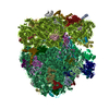
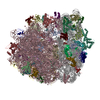
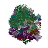
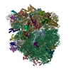
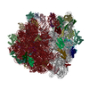
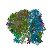
| Title | Visualization of two tRNAs trapped in transit during EF-G-mediated translocation | |||||||||
 Components Components |
| |||||||||
 Keywords Keywords | RIBOSOME/TRANSLATION / translocation intermediate / EF-G / head swiveling / RIBOSOME-TRANSLATION complex | |||||||||
| Function / homology |  Function and homology information Function and homology informationribosome disassembly / guanosine tetraphosphate binding / stringent response / ornithine decarboxylase inhibitor activity / transcription antitermination factor activity, RNA binding / misfolded RNA binding / Group I intron splicing / RNA folding / translational elongation / transcriptional attenuation ...ribosome disassembly / guanosine tetraphosphate binding / stringent response / ornithine decarboxylase inhibitor activity / transcription antitermination factor activity, RNA binding / misfolded RNA binding / Group I intron splicing / RNA folding / translational elongation / transcriptional attenuation / endoribonuclease inhibitor activity / RNA-binding transcription regulator activity / positive regulation of ribosome biogenesis / negative regulation of cytoplasmic translation / translation elongation factor activity / four-way junction DNA binding / translational termination / DnaA-L2 complex / translation repressor activity / negative regulation of DNA-templated DNA replication initiation / negative regulation of translational initiation / regulation of mRNA stability / mRNA regulatory element binding translation repressor activity / ribosome assembly / positive regulation of RNA splicing / assembly of large subunit precursor of preribosome / transcription elongation factor complex / regulation of DNA-templated transcription elongation / cytosolic ribosome assembly / DNA endonuclease activity / response to reactive oxygen species / transcription antitermination / regulation of cell growth / DNA-templated transcription termination / maintenance of translational fidelity / response to radiation / mRNA 5'-UTR binding / ribosomal small subunit biogenesis / large ribosomal subunit / small ribosomal subunit rRNA binding / Hydrolases; Acting on acid anhydrides; Acting on GTP to facilitate cellular and subcellular movement / ribosome biogenesis / ribosome binding / regulation of translation / ribosomal small subunit assembly / ribosomal large subunit assembly / small ribosomal subunit / transferase activity / large ribosomal subunit rRNA binding / 5S rRNA binding / cytosolic small ribosomal subunit / cytoplasmic translation / cytosolic large ribosomal subunit / tRNA binding / molecular adaptor activity / negative regulation of translation / rRNA binding / ribosome / structural constituent of ribosome / translation / response to antibiotic / negative regulation of DNA-templated transcription / GTPase activity / mRNA binding / GTP binding / protein homodimerization activity / DNA binding / RNA binding / zinc ion binding / membrane / cytosol / cytoplasm Similarity search - Function | |||||||||
| Biological species |  | |||||||||
| Method | ELECTRON MICROSCOPY / single particle reconstruction / cryo EM / Resolution: 6.8 Å | |||||||||
 Authors Authors | Ramrath, D.J.F. / Lancaster, L. / Sprink, T. / Mielke, T. / Loerke, J. / Noller, H.F. / Spahn, C.M.T. | |||||||||
 Citation Citation | Journal: Proc Natl Acad Sci U S A / Year: 2013 Title: Visualization of two transfer RNAs trapped in transit during elongation factor G-mediated translocation. Authors: David J F Ramrath / Laura Lancaster / Thiemo Sprink / Thorsten Mielke / Justus Loerke / Harry F Noller / Christian M T Spahn /  Abstract: During protein synthesis, coupled translocation of messenger RNAs (mRNA) and transfer RNAs (tRNA) through the ribosome takes place following formation of each peptide bond. The reaction is ...During protein synthesis, coupled translocation of messenger RNAs (mRNA) and transfer RNAs (tRNA) through the ribosome takes place following formation of each peptide bond. The reaction is facilitated by large-scale conformational changes within the ribosomal complex and catalyzed by elongtion factor G (EF-G). Previous structural analysis of the interaction of EF-G with the ribosome used either model complexes containing no tRNA or only a single tRNA, or complexes where EF-G was directly bound to ribosomes in the posttranslocational state. Here, we present a multiparticle cryo-EM reconstruction of a translocation intermediate containing two tRNAs trapped in transit, bound in chimeric intrasubunit ap/P and pe/E hybrid states. The downstream ap/P-tRNA is contacted by domain IV of EF-G and P-site elements within the 30S subunit body, whereas the upstream pe/E-tRNA maintains tight interactions with P-site elements of the swiveled 30S head. Remarkably, a tight compaction of the tRNA pair can be seen in this state. The translocational intermediate presented here represents a previously missing link in understanding the mechanism of translocation, revealing that the ribosome uses two distinct molecular ratchets, involving both intra- and intersubunit rotational movements, to drive the synchronous movement of tRNAs and mRNA. | |||||||||
| History |
|
- Structure visualization
| Movie |
Movie viewer |
|---|---|
| Structure viewer | Molecule: MolmilJmol/JSmol |
- Downloads & links
-Download
| PDBx/mmCIF format | 4v7b.cif.gz | 3.7 MB | Display | PDBx/mmCIF format |
|---|---|---|---|---|
| PDB format | pdb4v7b.ent.gz | Display | PDB format | |
| PDBx/mmJSON format | 4v7b.json.gz | Tree view | PDBx/mmJSON format | |
| Others |  Other downloads Other downloads |
-Validation report
| Summary document | 4v7b_validation.pdf.gz | 1.6 MB | Display | wwPDB validaton report |
|---|---|---|---|---|
| Full document | 4v7b_full_validation.pdf.gz | 2 MB | Display | |
| Data in XML | 4v7b_validation.xml.gz | 288.7 KB | Display | |
| Data in CIF | 4v7b_validation.cif.gz | 462.3 KB | Display | |
| Arichive directory | https://data.pdbj.org/pub/pdb/validation_reports/v7/4v7bftp://data.pdbj.org/pub/pdb/validation_reports/v7/4v7b | HTTPS FTP |
-Related structure data
| Related structure data | 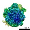 5775MC M: map data used to model this data C: citing same article ( |
|---|---|
| Similar structure data |


-Links
 PDBj
PDBj


- Assembly
| Deposited unit | 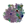
|
|---|---|
| 1 |
|
-Components
-RNA chain , 6 types, 6 molecules AAAVAWAXBBBA
| #1: RNA chain | Mass: 499690.031 Da / Num. of mol.: 1 / Source method: isolated from a natural source / Source: (natural) |
|---|---|
| #22: RNA chain | Mass: 24816.811 Da / Num. of mol.: 1 / Source method: isolated from a natural source / Source: (natural) |
| #23: RNA chain | Mass: 24728.689 Da / Num. of mol.: 1 / Source method: isolated from a natural source / Source: (natural) |
| #24: RNA chain | Mass: 6204.834 Da / Num. of mol.: 1 / Source method: isolated from a natural source / Source: (natural) |
| #26: RNA chain | Mass: 38790.090 Da / Num. of mol.: 1 / Source method: isolated from a natural source / Source: (natural) |
| #28: RNA chain | Mass: 941612.375 Da / Num. of mol.: 1 / Source method: isolated from a natural source / Source: (natural) |
-30S ribosomal protein ... , 20 types, 20 molecules ABACADAEAFAGAHAIAJAKALAMANAOAPAQARASATAU
| #2: Protein | Mass: 26781.670 Da / Num. of mol.: 1 / Source method: isolated from a natural source / Source: (natural) |
|---|---|
| #3: Protein | Mass: 26031.316 Da / Num. of mol.: 1 / Source method: isolated from a natural source / Source: (natural) |
| #4: Protein | Mass: 23514.199 Da / Num. of mol.: 1 / Source method: isolated from a natural source / Source: (natural) |
| #5: Protein | Mass: 17629.398 Da / Num. of mol.: 1 / Source method: isolated from a natural source / Source: (natural) |
| #6: Protein | Mass: 15727.512 Da / Num. of mol.: 1 / Source method: isolated from a natural source / Source: (natural) |
| #7: Protein | Mass: 20055.156 Da / Num. of mol.: 1 / Source method: isolated from a natural source / Source: (natural) |
| #8: Protein | Mass: 14146.557 Da / Num. of mol.: 1 / Source method: isolated from a natural source / Source: (natural) |
| #9: Protein | Mass: 14886.270 Da / Num. of mol.: 1 / Source method: isolated from a natural source / Source: (natural) |
| #10: Protein | Mass: 11755.597 Da / Num. of mol.: 1 / Source method: isolated from a natural source / Source: (natural) |
| #11: Protein | Mass: 13870.975 Da / Num. of mol.: 1 / Source method: isolated from a natural source / Source: (natural) |
| #12: Protein | Mass: 13768.157 Da / Num. of mol.: 1 / Source method: isolated from a natural source / Source: (natural) |
| #13: Protein | Mass: 13128.467 Da / Num. of mol.: 1 / Source method: isolated from a natural source / Source: (natural) |
| #14: Protein | Mass: 11606.560 Da / Num. of mol.: 1 / Source method: isolated from a natural source / Source: (natural) |
| #15: Protein | Mass: 10290.816 Da / Num. of mol.: 1 / Source method: isolated from a natural source / Source: (natural) |
| #16: Protein | Mass: 9207.572 Da / Num. of mol.: 1 / Source method: isolated from a natural source / Source: (natural) |
| #17: Protein | Mass: 9724.491 Da / Num. of mol.: 1 / Source method: isolated from a natural source / Source: (natural) |
| #18: Protein | Mass: 9005.472 Da / Num. of mol.: 1 / Source method: isolated from a natural source / Source: (natural) |
| #19: Protein | Mass: 10455.355 Da / Num. of mol.: 1 / Source method: isolated from a natural source / Source: (natural) |
| #20: Protein | Mass: 9708.464 Da / Num. of mol.: 1 / Source method: isolated from a natural source / Source: (natural) |
| #21: Protein | Mass: 8524.039 Da / Num. of mol.: 1 / Source method: isolated from a natural source / Source: (natural) |
-Protein , 1 types, 1 molecules AY
| #25: Protein | Mass: 77676.227 Da / Num. of mol.: 1 / Source method: isolated from a natural source / Source: (natural) |
|---|
+50S ribosomal protein ... , 31 types, 31 molecules BCBDBEBFBGBHBIBJBKBLBMBNBOBPBQBRBSBTBUBVBWBXBYBZB0B1B2B3B4B5B6
-Non-polymers , 2 types, 2 molecules 

| #59: Chemical | ChemComp-FUA /  Mass: 516.709 Da / Num. of mol.: 1 / Source method: obtained synthetically / Formula: C31H48O6 / Comment: antibiotic, Antimicrobial*YM Mass: 516.709 Da / Num. of mol.: 1 / Source method: obtained synthetically / Formula: C31H48O6 / Comment: antibiotic, Antimicrobial*YM |
|---|---|
| #60: Chemical | ChemComp-GDP /  Type: RNA linking / Mass: 443.201 Da / Num. of mol.: 1 / Source method: obtained synthetically / Formula: C10H15N5O11P2 / Comment: GDP, energy-carrying molecule*YM Type: RNA linking / Mass: 443.201 Da / Num. of mol.: 1 / Source method: obtained synthetically / Formula: C10H15N5O11P2 / Comment: GDP, energy-carrying molecule*YM |
-Experimental details
-Experiment
| Experiment | Method: ELECTRON MICROSCOPY |
|---|---|
| EM experiment | Aggregation state: PARTICLE / 3D reconstruction method: single particle reconstruction |
- Sample preparation
| Component | Name: ribosomal 70S-EF-G complex / Type: RIBOSOME / Details: in vitro reconstituted ribosomal complex, monomer |
|---|---|
| Molecular weight | Value: 3 MDa / Experimental value: YES |
| Buffer solution | Name: 80 mM HEPES potassium, 75 mM NH4Cl, 10 mM MgCl2, 6 mM BME pH: 7.6 Details: 80 mM HEPES potassium, 75 mM NH4Cl, 10 mM MgCl2, 6 mM BME |
| Specimen | Conc.: 0.15 mg/ml / Embedding applied: NO / Shadowing applied: NO / Staining applied: NO / Vitrification applied: YES |
| Specimen support | Details: Quantifoil R3-3 Cu mesh with 2 nm carbon support film |
| Vitrification | Instrument: FEI VITROBOT MARK I / Cryogen name: ETHANE / Temp: 96 K / Humidity: 100 % Details: Blot for 5-10 seconds before plunging into liquid ethane (FEI VITROBOT MARK I) Method: blot for 5-10 seconds before plunging |
- Electron microscopy imaging
| Experimental equipment |  Model: Tecnai Polara / Image courtesy: FEI Company |
|---|---|
| Microscopy | Model: FEI POLARA 300 / Date: Jun 14, 2010 |
| Electron gun | Electron source:  FIELD EMISSION GUN / Accelerating voltage: 300 kV / Illumination mode: SPOT SCAN FIELD EMISSION GUN / Accelerating voltage: 300 kV / Illumination mode: SPOT SCAN |
| Electron lens | Mode: BRIGHT FIELD / Nominal magnification: 39000 X / Calibrated magnification: 39000 X / Nominal defocus max: 5000 nm / Nominal defocus min: 2000 nm / Camera length: 0 mm |
| Specimen holder | Specimen holder model: GATAN HELIUM / Temperature: 77 K / Tilt angle max: 0 ° / Tilt angle min: 0 ° |
| Image recording | Electron dose: 20 e/Å2 / Film or detector model: KODAK SO-163 FILM |
| Image scans | Num. digital images: 308 |
| Radiation | Protocol: SINGLE WAVELENGTH / Monochromatic (M) / Laue (L): M / Scattering type: x-ray |
| Radiation wavelength | Relative weight: 1 |
- Processing
| EM software |
| ||||||||||||
|---|---|---|---|---|---|---|---|---|---|---|---|---|---|
| CTF correction | Details: The volumes were CTF-corrected in defocus groups with an average of approximately 906 individual images per group. | ||||||||||||
| Symmetry | Point symmetry: C1 (asymmetric) | ||||||||||||
| 3D reconstruction | Method: angular refinement / Resolution: 6.8 Å / Resolution method: FSC 0.5 CUT-OFF / Num. of particles: 279309 / Nominal pixel size: 1.26 Å / Actual pixel size: 1.26 Å / Details: (Single particle--Applied symmetry: C1) / Symmetry type: POINT | ||||||||||||
| Atomic model building | Protocol: RIGID BODY FIT / Space: REAL / Details: REFINEMENT PROTOCOL--rigid body | ||||||||||||
| Atomic model building | PDB-ID: 4KIY 4kiy | ||||||||||||
| Refinement | Highest resolution: 6.8 Å | ||||||||||||
| Refinement step | Cycle: LAST / Highest resolution: 6.8 Å
|
