Movie
Movie Controller

[English] 日本語
 Yorodumi
- PDB-4uxy: Conserved mechanisms of microtubule-stimulated ADP release, ATP b... -
+ Open data

- Basic information
| Entry | Database: PDB / ID: 4uxy | ||||||
|---|---|---|---|---|---|---|---|
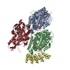
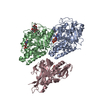
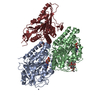
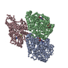
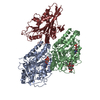
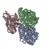
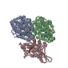
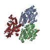
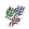
| Title | Conserved mechanisms of microtubule-stimulated ADP release, ATP binding, and force generation in transport kinesins | ||||||
 Components Components |
| ||||||
 Keywords Keywords | TRANSPORT PROTEIN / KINESIN / MICROTUBULE / CRYO-EM | ||||||
| Function / homology |  Function and homology information Function and homology informationplus-end-directed kinesin ATPase / anterograde dendritic transport of neurotransmitter receptor complex / anterograde axonal protein transport / retrograde neuronal dense core vesicle transport / ciliary rootlet / plus-end-directed microtubule motor activity / RHO GTPases activate KTN1 / Kinesins / positive regulation of axon guidance / microtubule motor activity ...plus-end-directed kinesin ATPase / anterograde dendritic transport of neurotransmitter receptor complex / anterograde axonal protein transport / retrograde neuronal dense core vesicle transport / ciliary rootlet / plus-end-directed microtubule motor activity / RHO GTPases activate KTN1 / Kinesins / positive regulation of axon guidance / microtubule motor activity / kinesin complex / COPI-dependent Golgi-to-ER retrograde traffic / microtubule-based movement / Insulin processing / synaptic vesicle transport / cytoskeletal motor activity / kinesin binding / postsynaptic cytosol / microtubule-based process / cytoplasmic microtubule / vesicle-mediated transport / axon cytoplasm / MHC class II antigen presentation / cellular response to interleukin-4 / dendrite cytoplasm / axon guidance / structural constituent of cytoskeleton / microtubule cytoskeleton organization / neuron migration / mitotic cell cycle / double-stranded RNA binding / microtubule cytoskeleton / microtubule binding / chemical synaptic transmission / Hydrolases; Acting on acid anhydrides; Acting on GTP to facilitate cellular and subcellular movement / microtubule / perikaryon / cilium / protein heterodimerization activity / GTPase activity / ubiquitin protein ligase binding / GTP binding / perinuclear region of cytoplasm / ATP hydrolysis activity / ATP binding / membrane / metal ion binding / cytoplasm / cytosol Similarity search - Function | ||||||
| Biological species |  HOMO SAPIENS (human) HOMO SAPIENS (human) | ||||||
| Method | ELECTRON MICROSCOPY / single particle reconstruction / cryo EM / Resolution: 6.5 Å | ||||||
| Model type details | CA ATOMS ONLY, CHAIN A, B, C | ||||||
 Authors Authors | Atherton, J. / Farabella, I. / Yu, I.M. / Rosenfeld, S.S. / Houdusse, A. / Topf, M. / Moores, C. | ||||||
 Citation Citation | Journal: Elife / Year: 2014 Title: Conserved mechanisms of microtubule-stimulated ADP release, ATP binding, and force generation in transport kinesins. Authors: Joseph Atherton / Irene Farabella / I-Mei Yu / Steven S Rosenfeld / Anne Houdusse / Maya Topf / Carolyn A Moores /    Abstract: Kinesins are a superfamily of microtubule-based ATP-powered motors, important for multiple, essential cellular functions. How microtubule binding stimulates their ATPase and controls force generation ...Kinesins are a superfamily of microtubule-based ATP-powered motors, important for multiple, essential cellular functions. How microtubule binding stimulates their ATPase and controls force generation is not understood. To address this fundamental question, we visualized microtubule-bound kinesin-1 and kinesin-3 motor domains at multiple steps in their ATPase cycles--including their nucleotide-free states--at ∼ 7 Å resolution using cryo-electron microscopy. In both motors, microtubule binding promotes ordered conformations of conserved loops that stimulate ADP release, enhance microtubule affinity and prime the catalytic site for ATP binding. ATP binding causes only small shifts of these nucleotide-coordinating loops but induces large conformational changes elsewhere that allow force generation and neck linker docking towards the microtubule plus end. Family-specific differences across the kinesin-microtubule interface account for the distinctive properties of each motor. Our data thus provide evidence for a conserved ATP-driven mechanism for kinesins and reveal the critical mechanistic contribution of the microtubule interface. | ||||||
| History |
|
- Structure visualization
| Movie |
Movie viewer |
|---|---|
| Structure viewer | Molecule: MolmilJmol/JSmol |
- Downloads & links
-Download
| PDBx/mmCIF format | 4uxy.cif.gz | 65.3 KB | Display | PDBx/mmCIF format |
|---|---|---|---|---|
| PDB format | pdb4uxy.ent.gz | 34.2 KB | Display | PDB format |
| PDBx/mmJSON format | 4uxy.json.gz | Tree view | PDBx/mmJSON format | |
| Others |  Other downloads Other downloads |
-Validation report
| Arichive directory | https://data.pdbj.org/pub/pdb/validation_reports/ux/4uxyftp://data.pdbj.org/pub/pdb/validation_reports/ux/4uxy | HTTPS FTP |
|---|
-Related structure data
| Related structure data |  2770MC 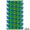 2765C 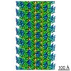 2766C 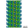 2767C  2768C  2769C  2771C  4uxoC  4uxpC  4uxrC  4uxsC  4uxtC  4uy0C M: map data used to model this data C: citing same article ( |
|---|---|
| Similar structure data |


-Links
 PDBj
PDBj


- Assembly
| Deposited unit | 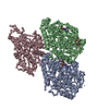
|
|---|---|
| 1 |
|
-Components
-Protein , 3 types, 3 molecules ABC
| #1: Protein | Mass: 50107.238 Da / Num. of mol.: 1 / Source method: isolated from a natural source / Source: (natural) |
|---|---|
| #2: Protein | Mass: 49907.770 Da / Num. of mol.: 1 / Source method: isolated from a natural source / Source: (natural) |
| #3: Protein | Mass: 38030.184 Da / Num. of mol.: 1 Source method: isolated from a genetically manipulated source Details: WITH BOUND AMPPNP / Source: (gene. exp.) HOMO SAPIENS (human) / Plasmid: PET151-D-TOPO / Production host:  |
-Non-polymers , 5 types, 5 molecules 


| #4: Chemical | ChemComp-ZN /  Mass: 65.409 Da / Num. of mol.: 1 / Source method: obtained synthetically / Formula: Zn Mass: 65.409 Da / Num. of mol.: 1 / Source method: obtained synthetically / Formula: Zn |
|---|---|
| #5: Chemical | ChemComp-MG /  Mass: 24.305 Da / Num. of mol.: 1 / Source method: obtained synthetically / Formula: Mg Mass: 24.305 Da / Num. of mol.: 1 / Source method: obtained synthetically / Formula: Mg |
| #6: Chemical | ChemComp-GTP /  Mass: 523.180 Da / Num. of mol.: 1 / Source method: obtained synthetically / Formula: C10H16N5O14P3 / Comment: GTP, energy-carrying molecule*YM Mass: 523.180 Da / Num. of mol.: 1 / Source method: obtained synthetically / Formula: C10H16N5O14P3 / Comment: GTP, energy-carrying molecule*YM |
| #7: Chemical | ChemComp-GDP /  Type: RNA linking / Mass: 443.201 Da / Num. of mol.: 1 / Source method: obtained synthetically / Formula: C10H15N5O11P2 / Comment: GDP, energy-carrying molecule*YM Type: RNA linking / Mass: 443.201 Da / Num. of mol.: 1 / Source method: obtained synthetically / Formula: C10H15N5O11P2 / Comment: GDP, energy-carrying molecule*YM |
| #8: Chemical | ChemComp-TA1 /  Mass: 853.906 Da / Num. of mol.: 1 / Source method: obtained synthetically / Formula: C47H51NO14 / Comment: medication, chemotherapy*YM Mass: 853.906 Da / Num. of mol.: 1 / Source method: obtained synthetically / Formula: C47H51NO14 / Comment: medication, chemotherapy*YM |
-Experimental details
-Experiment
| Experiment | Method: ELECTRON MICROSCOPY |
|---|---|
| EM experiment | Aggregation state: HELICAL ARRAY / 3D reconstruction method: single particle reconstruction |
- Sample preparation
| Component | Name: 3-protofilament microtubule-bound human kinesin-1 motor domain with AMPPNP Type: COMPLEX |
|---|---|
| Buffer solution | pH: 6.8 |
| Specimen | Embedding applied: NO / Shadowing applied: NO / Staining applied: NO / Vitrification applied: YES |
| Vitrification | Instrument: FEI VITROBOT MARK I / Cryogen name: ETHANE |
- Electron microscopy imaging
| Experimental equipment |  Model: Tecnai Polara / Image courtesy: FEI Company |
|---|---|
| Microscopy | Model: FEI POLARA 300 / Date: Dec 10, 2012 |
| Electron gun | Electron source:  FIELD EMISSION GUN / Accelerating voltage: 300 kV / Illumination mode: FLOOD BEAM FIELD EMISSION GUN / Accelerating voltage: 300 kV / Illumination mode: FLOOD BEAM |
| Electron lens | Mode: BRIGHT FIELD / Calibrated magnification: 100000 X / Nominal defocus max: 3500 nm / Nominal defocus min: 400 nm / Cs: 2.3 mm |
| Specimen holder | Temperature: 90 K |
| Image recording | Electron dose: 20 e/Å2 / Film or detector model: GATAN ULTRASCAN 4000 (4k x 4k) |
| Image scans | Num. digital images: 519 |
| Radiation wavelength | Relative weight: 1 |
- Processing
| EM software |
| |||||||||||||||
|---|---|---|---|---|---|---|---|---|---|---|---|---|---|---|---|---|
| CTF correction | Details: FREALIGN | |||||||||||||||
| 3D reconstruction | Resolution: 6.5 Å / Num. of particles: 186329 / Actual pixel size: 1.5 Å Details: SUBMISSION BASED ON EXPERIMENTAL DATA FROM EMDB EMD-2770. (DEPOSITION ID: 12603). Symmetry type: POINT | |||||||||||||||
| Atomic model building | Protocol: FLEXIBLE FIT / Space: REAL / Target criteria: Cross-correlation coefficient / Details: METHOD--FLEXIBLE REFINEMENT PROTOCOL--X-RAY | |||||||||||||||
| Atomic model building | PDB-ID: 4HNA Accession code: 4HNA / Source name: PDB / Type: experimental model | |||||||||||||||
| Refinement | Highest resolution: 6.5 Å | |||||||||||||||
| Refinement step | Cycle: LAST / Highest resolution: 6.5 Å
|