Movie
Movie Controller

[English] 日本語
 Yorodumi
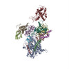
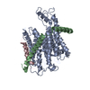
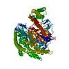
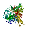
- PDB-4cg6: Cryo-em of the Sec61-complex bound to the 80s ribosome translatin... -
+ Open data

- Basic information
| Entry | Database: PDB / ID: 4cg6 | ||||||
|---|---|---|---|---|---|---|---|
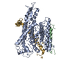
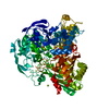
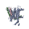
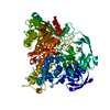
| Title | Cryo-em of the Sec61-complex bound to the 80s ribosome translating a membrane-inserting substrate | ||||||
 Components Components |
| ||||||
 Keywords Keywords | PROTEIN TRANSPORT / CO-TRANSLATIONAL PROTEIN TRANSLOCATION | ||||||
| Function / homology |  Function and homology information Function and homology informationInsertion of tail-anchored proteins into the endoplasmic reticulum membrane / membrane docking / endoplasmic reticulum Sec complex / pronephric nephron development / cotranslational protein targeting to membrane / Ssh1 translocon complex / Sec61 translocon complex / protein insertion into ER membrane / protein-transporting ATPase activity / protein targeting to ER ...Insertion of tail-anchored proteins into the endoplasmic reticulum membrane / membrane docking / endoplasmic reticulum Sec complex / pronephric nephron development / cotranslational protein targeting to membrane / Ssh1 translocon complex / Sec61 translocon complex / protein insertion into ER membrane / protein-transporting ATPase activity / protein targeting to ER / SRP-dependent cotranslational protein targeting to membrane, translocation / signal sequence receptor activity / post-translational protein targeting to membrane, translocation / transmembrane protein transporter activity / guanyl-nucleotide exchange factor activity / phospholipid binding / ribosome binding / endoplasmic reticulum membrane / endoplasmic reticulum / membrane Similarity search - Function | ||||||
| Biological species |  | ||||||
| Method | ELECTRON MICROSCOPY / single particle reconstruction / cryo EM / Resolution: 7.8 Å | ||||||
 Authors Authors | Gogala, M. / Becker, T. / Beatrix, B. / Barrio-Garcia, C. / Berninghausen, O. / Beckmann, R. | ||||||
 Citation Citation | Journal: Nature / Year: 2014 Title: Structures of the Sec61 complex engaged in nascent peptide translocation or membrane insertion. Authors: Marko Gogala / Thomas Becker / Birgitta Beatrix / Jean-Paul Armache / Clara Barrio-Garcia / Otto Berninghausen / Roland Beckmann /  Abstract: The biogenesis of secretory as well as transmembrane proteins requires the activity of the universally conserved protein-conducting channel (PCC), the Sec61 complex (SecY complex in bacteria). In ...The biogenesis of secretory as well as transmembrane proteins requires the activity of the universally conserved protein-conducting channel (PCC), the Sec61 complex (SecY complex in bacteria). In eukaryotic cells the PCC is located in the membrane of the endoplasmic reticulum where it can bind to translating ribosomes for co-translational protein transport. The Sec complex consists of three subunits (Sec61α, β and γ) and provides an aqueous environment for the translocation of hydrophilic peptides as well as a lateral opening in the Sec61α subunit that has been proposed to act as a gate for the membrane partitioning of hydrophobic domains. A plug helix and a so-called pore ring are believed to seal the PCC against ion flow and are proposed to rearrange for accommodation of translocating peptides. Several crystal and cryo-electron microscopy structures revealed different conformations of closed and partially open Sec61 and SecY complexes. However, in none of these samples has the translocation state been unambiguously defined biochemically. Here we present cryo-electron microscopy structures of ribosome-bound Sec61 complexes engaged in translocation or membrane insertion of nascent peptides. Our data show that a hydrophilic peptide can translocate through the Sec complex with an essentially closed lateral gate and an only slightly rearranged central channel. Membrane insertion of a hydrophobic domain seems to occur with the Sec complex opening the proposed lateral gate while rearranging the plug to maintain an ion permeability barrier. Taken together, we provide a structural model for the basic activities of the Sec61 complex as a protein-conducting channel. | ||||||
| History |
|
- Structure visualization
| Movie |
Movie viewer |
|---|---|
| Structure viewer | Molecule: MolmilJmol/JSmol |
- Downloads & links
-Download
| PDBx/mmCIF format | 4cg6.cif.gz | 107.5 KB | Display | PDBx/mmCIF format |
|---|---|---|---|---|
| PDB format | pdb4cg6.ent.gz | 79.7 KB | Display | PDB format |
| PDBx/mmJSON format | 4cg6.json.gz | Tree view | PDBx/mmJSON format | |
| Others |  Other downloads Other downloads |
-Validation report
| Arichive directory | https://data.pdbj.org/pub/pdb/validation_reports/cg/4cg6ftp://data.pdbj.org/pub/pdb/validation_reports/cg/4cg6 | HTTPS FTP |
|---|
-Related structure data
| Related structure data | 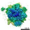 2512MC 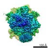 2510C 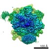 2511C  4cg5C 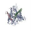 4cg7C  4v7eC M: map data used to model this data C: citing same article ( |
|---|---|
| Similar structure data |
-Links
 PDBj
PDBj

- Assembly
| Deposited unit | 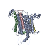
|
|---|---|
| 1 |
|
-Components
| #1: Protein | Mass: 52279.379 Da / Num. of mol.: 1 / Source method: isolated from a natural source / Details: DATA-SUBSET RESULTED FROM COMPUTATIONAL SORTING / Source: (natural) |
|---|---|
| #2: Protein | Mass: 7752.325 Da / Num. of mol.: 1 / Source method: isolated from a natural source / Details: DATA-SUBSET RESULTED FROM COMPUTATIONAL SORTING / Source: (natural) |
| #3: Protein | Mass: 9987.456 Da / Num. of mol.: 1 / Source method: isolated from a natural source / Details: DATA-SUBSET RESULTED FROM COMPUTATIONAL SORTING / Source: (natural) |
| #4: Protein/peptide | Mass: 1839.265 Da / Num. of mol.: 1 / Source method: isolated from a natural source / Details: DATA-SUBSET RESULTED FROM COMPUTATIONAL SORTING / Source: (natural) |
-Experimental details
-Experiment
| Experiment | Method: ELECTRON MICROSCOPY |
|---|---|
| EM experiment | Aggregation state: PARTICLE / 3D reconstruction method: single particle reconstruction |
- Sample preparation
| Component | Name: CANIS FAMILIARIS SEC61 BOUND TO A WHEAT GERM 80S-RNC TRANSLATING THE MEMBRANE- INSERTING LEPM-POLYPEPTIDE Type: RIBOSOME |
|---|---|
| Buffer solution | Name: 30 MM HEPES/KOH 7.6, 10 MM MG(OAC)2, 180 MM KOAC/HAC PH 7.6, 0.3 % DIGITONIN, 1 MM DTT pH: 7.6 Details: 30 MM HEPES/KOH 7.6, 10 MM MG(OAC)2, 180 MM KOAC/HAC PH 7.6, 0.3 % DIGITONIN, 1 MM DTT |
| Specimen | Embedding applied: NO / Shadowing applied: NO / Staining applied: NO / Vitrification applied: YES |
| Specimen support | Details: CARBON |
| Vitrification | Instrument: FEI VITROBOT MARK IV / Cryogen name: ETHANE Details: VITRIFICATION 1 -- CRYOGEN- ETHANE, HUMIDITY- 95, INSTRUMENT- FEI VITROBOT MARK IV, METHOD- BLOT FOR 3 SECONDS BEFORE PLUNGING, |
- Electron microscopy imaging
| Experimental equipment |  Model: Titan Krios / Image courtesy: FEI Company |
|---|---|
| Microscopy | Model: FEI TITAN KRIOS / Date: Jul 17, 2011 |
| Electron gun | Electron source:  FIELD EMISSION GUN / Accelerating voltage: 200 kV / Illumination mode: FLOOD BEAM FIELD EMISSION GUN / Accelerating voltage: 200 kV / Illumination mode: FLOOD BEAM |
| Electron lens | Mode: BRIGHT FIELD / Calibrated magnification: 148721 X / Nominal defocus max: 4000 nm / Nominal defocus min: 1300 nm / Cs: 2.7 mm |
| Image recording | Electron dose: 25 e/Å2 / Film or detector model: TVIPS TEMCAM-F416 (4k x 4k) |
| Image scans | Num. digital images: 9805 |
| Radiation wavelength | Relative weight: 1 |
- Processing
| EM software |
| |||||||||||||||||||||
|---|---|---|---|---|---|---|---|---|---|---|---|---|---|---|---|---|---|---|---|---|---|---|
| CTF correction | Details: ON 3D-VOLUME (SPIDER) | |||||||||||||||||||||
| Symmetry | Point symmetry: C1 (asymmetric) | |||||||||||||||||||||
| 3D reconstruction | Resolution: 7.8 Å / Num. of particles: 30455 Details: SUBMISSION BASED ON EXPERIMENTAL DATA FROM EMDB EMD-2512 (DEPOSITION ID: 12127). Symmetry type: POINT | |||||||||||||||||||||
| Atomic model building | Protocol: FLEXIBLE FIT / Space: REAL / Details: METHOD--FLEXIBLE | |||||||||||||||||||||
| Atomic model building | PDB-ID: 2WWB Accession code: 2WWB / Source name: PDB / Type: experimental model | |||||||||||||||||||||
| Refinement | Highest resolution: 7.8 Å | |||||||||||||||||||||
| Refinement step | Cycle: LAST / Highest resolution: 7.8 Å
|