 Movie
Movie Controller

[English] 日本語
 Yorodumi
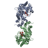
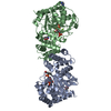
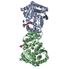
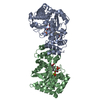
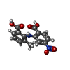
- PDB-4zof: Lobenzarit-like inhibitor bound in the active site of Mycobacteri... -
+ Open data

- Basic information
| Entry | Database: PDB / ID: 4zof | |||||||||
|---|---|---|---|---|---|---|---|---|---|---|
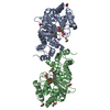
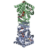
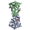
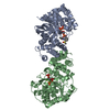
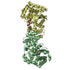
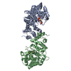
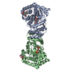
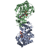
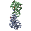
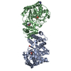
| Title | Lobenzarit-like inhibitor bound in the active site of Mycobacterium tuberculosis anthranilate phosphoribosyltransferase (AnPRT; trpD) | |||||||||
 Components Components | Anthranilate phosphoribosyltransferase | |||||||||
 Keywords Keywords | TRANSFERASE/TRANSFERASE INHIBITOR / inhibitor / complex / Lobenzarit-like analogue / TB Structural Genomics Consortium / TBSGC / phosphoribosyltransferase / magnesium binding / phosphoribosylpyrophosphate / PRPP / TRANSFERASE-TRANSFERASE INHIBITOR complex | |||||||||
| Function / homology |  Function and homology information Function and homology informationanthranilate phosphoribosyltransferase / anthranilate phosphoribosyltransferase activity / L-tryptophan biosynthetic process / magnesium ion binding / cytosol Similarity search - Function | |||||||||
| Biological species |  Mycobacterium tuberculosis H37Ra (bacteria) Mycobacterium tuberculosis H37Ra (bacteria) | |||||||||
| Method |  X-RAY DIFFRACTION / SYNCHROTRON / MOLECULAR REPLACEMENT / Resolution: 1.8 Å X-RAY DIFFRACTION / SYNCHROTRON / MOLECULAR REPLACEMENT / Resolution: 1.8 Å | |||||||||
 Authors Authors | Evans, G.L. / Baker, E.N. / Lott, J.S. / TB Structural Genomics Consortium (TBSGC) | |||||||||
| Funding support |  New Zealand, 2items New Zealand, 2items
| |||||||||
 Citation Citation | Journal: To be Published Title: Inhibitor bound in the active site of Mycobacterium tuberculosis anthranilate phosphoribosyltransferase (AnPRT; trpD). Authors: Evans, G.L. / Baker, E.N. / Lott, J.S. | |||||||||
| History |
|
- Structure visualization
| Structure viewer | Molecule: MolmilJmol/JSmol |
|---|
- Downloads & links
-Download
| PDBx/mmCIF format | 4zof.cif.gz | 150.9 KB | Display | PDBx/mmCIF format |
|---|---|---|---|---|
| PDB format | pdb4zof.ent.gz | 115 KB | Display | PDB format |
| PDBx/mmJSON format | 4zof.json.gz | Tree view | PDBx/mmJSON format | |
| Others |  Other downloads Other downloads |
-Validation report
| Arichive directory | https://data.pdbj.org/pub/pdb/validation_reports/zo/4zofftp://data.pdbj.org/pub/pdb/validation_reports/zo/4zof | HTTPS FTP |
|---|
-Related structure data
| Related structure data | 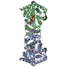 4zojC  4zokC 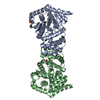 3qr9S S: Starting model for refinement C: citing same article ( |
|---|---|
| Similar structure data |


-Links
 PDBj
PDBj


- Assembly
| Deposited unit | 
| ||||||||
|---|---|---|---|---|---|---|---|---|---|
| 1 |
| ||||||||
| Unit cell |
|
-Components
-Protein / Sugars , 2 types, 4 molecules AB
| #1: Protein | Mass: 38948.012 Da / Num. of mol.: 2 Source method: isolated from a genetically manipulated source Source: (gene. exp.) Mycobacterium tuberculosis H37Ra (bacteria)Strain: ATCC 25177 / H37Ra / Gene: trpD, MRA_2208 / Plasmid: pET23A / Production host: References: UniProt: A5U4M0, anthranilate phosphoribosyltransferase #3: Sugar |  Type: D-saccharide / Mass: 390.070 Da / Num. of mol.: 2 / Source method: obtained synthetically / Formula: C5H13O14P3 Type: D-saccharide / Mass: 390.070 Da / Num. of mol.: 2 / Source method: obtained synthetically / Formula: C5H13O14P3 |
|---|
-Non-polymers , 5 types, 411 molecules 


| #2: Chemical | ChemComp-MG /  Mass: 24.305 Da / Num. of mol.: 4 / Source method: obtained synthetically / Formula: Mg Mass: 24.305 Da / Num. of mol.: 4 / Source method: obtained synthetically / Formula: Mg#4: Chemical |  Mass: 316.266 Da / Num. of mol.: 2 / Source method: obtained synthetically / Formula: C15H12N2O6 Mass: 316.266 Da / Num. of mol.: 2 / Source method: obtained synthetically / Formula: C15H12N2O6#5: Chemical | ChemComp-IMD / |  Mass: 69.085 Da / Num. of mol.: 1 / Source method: obtained synthetically / Formula: C3H5N2 Mass: 69.085 Da / Num. of mol.: 1 / Source method: obtained synthetically / Formula: C3H5N2#6: Chemical | ChemComp-PO4 / |  Mass: 94.971 Da / Num. of mol.: 1 / Source method: obtained synthetically / Formula: PO4 Mass: 94.971 Da / Num. of mol.: 1 / Source method: obtained synthetically / Formula: PO4#7: Water | ChemComp-HOH / | Mass: 18.015 Da / Num. of mol.: 403 / Source method: isolated from a natural source / Formula: H2O |
|---|
-Experimental details
-Experiment
| Experiment | Method: X-RAY DIFFRACTION |
|---|
- Sample preparation
| Crystal | Density Matthews: 2.27 Å3/Da / Density % sol: 45.9 % / Description: Flat diamond shape |
|---|---|
| Crystal grow | Temperature: 291 K / Method: vapor diffusion, hanging drop / pH: 7.5 / Details: 0.2 M imidazole-malate, 15% PEG4000 |
-Data collection
| Diffraction | Mean temperature: 100 K |
|---|---|
| Diffraction source | Source: SYNCHROTRON / Site: Australian Synchrotron  / Beamline: MX1 / Wavelength: 0.9537 Å / Beamline: MX1 / Wavelength: 0.9537 Å |
| Detector | Type: ADSC QUANTUM 210r / Detector: CCD / Date: Oct 30, 2014 |
| Radiation | Monochromator: double crystal Si(111) / Protocol: SINGLE WAVELENGTH / Monochromatic (M) / Laue (L): M / Scattering type: x-ray |
| Radiation wavelength | Wavelength: 0.9537 Å / Relative weight: 1 |
| Reflection | Resolution: 1.8→47 Å / Num. obs: 63649 / % possible obs: 100 % / Observed criterion σ(I): -3 / Redundancy: 7.5 % / Rmerge(I) obs: 0.179 / Net I/σ(I): 7.3 |
- Processing
| Software |
| |||||||||||||||||||||||||||||||||||||||||||||||||||||||||||||||||||||||||||||||||||||||||||||||||||||||||||||||||||||||
|---|---|---|---|---|---|---|---|---|---|---|---|---|---|---|---|---|---|---|---|---|---|---|---|---|---|---|---|---|---|---|---|---|---|---|---|---|---|---|---|---|---|---|---|---|---|---|---|---|---|---|---|---|---|---|---|---|---|---|---|---|---|---|---|---|---|---|---|---|---|---|---|---|---|---|---|---|---|---|---|---|---|---|---|---|---|---|---|---|---|---|---|---|---|---|---|---|---|---|---|---|---|---|---|---|---|---|---|---|---|---|---|---|---|---|---|---|---|---|---|---|
| Refinement | Method to determine structure: MOLECULAR REPLACEMENT Starting model: PDB entry 3QR9 chain A Resolution: 1.8→35.809 Å / SU ML: 0.21 / Cross valid method: FREE R-VALUE / σ(F): 1.33 / Phase error: 21.95 / Stereochemistry target values: ML
| |||||||||||||||||||||||||||||||||||||||||||||||||||||||||||||||||||||||||||||||||||||||||||||||||||||||||||||||||||||||
| Solvent computation | Shrinkage radii: 0.9 Å / VDW probe radii: 1.11 Å / Solvent model: FLAT BULK SOLVENT MODEL | |||||||||||||||||||||||||||||||||||||||||||||||||||||||||||||||||||||||||||||||||||||||||||||||||||||||||||||||||||||||
| Refinement step | Cycle: LAST / Resolution: 1.8→35.809 Å
| |||||||||||||||||||||||||||||||||||||||||||||||||||||||||||||||||||||||||||||||||||||||||||||||||||||||||||||||||||||||
| Refine LS restraints |
| |||||||||||||||||||||||||||||||||||||||||||||||||||||||||||||||||||||||||||||||||||||||||||||||||||||||||||||||||||||||
| LS refinement shell |
|
