cellular response to vitamin B1 / response to formaldehyde / response to water-immersion restraint stress / response to ether / traversing start control point of mitotic cell cycle / atrial septum development / fibroblast activation / regulation of protein catabolic process at postsynapse, modulating synaptic transmission / Trafficking of AMPA receptors / negative regulation of intrinsic apoptotic signaling pathway by p53 class mediator ...cellular response to vitamin B1 / response to formaldehyde / response to water-immersion restraint stress / response to ether / traversing start control point of mitotic cell cycle / atrial septum development / fibroblast activation / regulation of protein catabolic process at postsynapse, modulating synaptic transmission / Trafficking of AMPA receptors / negative regulation of intrinsic apoptotic signaling pathway by p53 class mediator / receptor serine/threonine kinase binding / negative regulation of protein processing / response to steroid hormone / SUMO transferase activity / peroxisome proliferator activated receptor binding / positive regulation of vascular associated smooth muscle cell migration / atrioventricular valve morphogenesis / AKT phosphorylates targets in the cytosol / response to iron ion / NEDD8 ligase activity / endocardial cushion morphogenesis / ventricular septum development / cellular response to peptide hormone stimulus / positive regulation of muscle cell differentiation / cardiac septum morphogenesis / regulation of postsynaptic neurotransmitter receptor internalization / SUMOylation of ubiquitinylation proteins / cellular response to alkaloid / blood vessel development / ligase activity / Constitutive Signaling by AKT1 E17K in Cancer / negative regulation of DNA damage response, signal transduction by p53 class mediator / cellular response to antibiotic / SUMOylation of transcription factors / negative regulation of signal transduction by p53 class mediator / regulation of protein catabolic process / cellular response to UV-C / cellular response to estrogen stimulus / response to magnesium ion / protein sumoylation / blood vessel remodeling / ribonucleoprotein complex binding / protein localization to nucleus / protein autoubiquitination / positive regulation of vascular associated smooth muscle cell proliferation / NPAS4 regulates expression of target genes / transcription repressor complex / positive regulation of mitotic cell cycle / regulation of heart rate / : / positive regulation of protein export from nucleus / response to cocaine / ubiquitin binding / DNA damage response, signal transduction by p53 class mediator / establishment of protein localization / Stabilization of p53 / sperm end piece / cellular response to gamma radiation / Regulation of RUNX3 expression and activity / RING-type E3 ubiquitin transferase / Oncogene Induced Senescence / Regulation of TP53 Activity through Methylation / protein destabilization / cellular response to growth factor stimulus / Degradation of CDH1 / response to toxic substance / cellular response to hydrogen peroxide / centriolar satellite / disordered domain specific binding / protein polyubiquitination / p53 binding / ubiquitin-protein transferase activity / endocytic vesicle membrane / Signaling by ALK fusions and activated point mutants / Regulation of TP53 Degradation / ubiquitin protein ligase activity / negative regulation of neuron projection development / positive regulation of proteasomal ubiquitin-dependent protein catabolic process / sperm principal piece / 5S rRNA binding / protein-containing complex assembly / sperm midpiece / Oxidative Stress Induced Senescence / cellular response to hypoxia / Regulation of TP53 Activity through Phosphorylation / amyloid fibril formation / ubiquitin-dependent protein catabolic process / proteasome-mediated ubiquitin-dependent protein catabolic process / regulation of cell cycle / postsynaptic density / Ub-specific processing proteases / protein ubiquitination / response to xenobiotic stimulus / protein domain specific binding / response to antibiotic / negative regulation of DNA-templated transcription / apoptotic process / positive regulation of cell population proliferation / ubiquitin protein ligase binding / positive regulation of gene expression Similarity search - Function
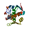
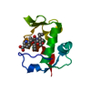
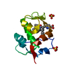
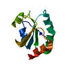
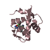
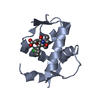
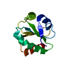
E3 ubiquitin-protein ligase Mdm2 / MDM2, modified RING finger, HC subclass / MDM2 / SWIB/MDM2 domain / p53 negative regulator Mdm2/Mdm4 / SWIB/MDM2 domain / SWIB/MDM2 domain / SWIB/MDM2 domain profile. / SWIB/MDM2 domain superfamily / Zn-finger in Ran binding protein and others ...E3 ubiquitin-protein ligase Mdm2 / MDM2, modified RING finger, HC subclass / MDM2 / SWIB/MDM2 domain / p53 negative regulator Mdm2/Mdm4 / SWIB/MDM2 domain / SWIB/MDM2 domain / SWIB/MDM2 domain profile. / SWIB/MDM2 domain superfamily / Zn-finger in Ran binding protein and others / Zinc finger, C3HC4 type (RING finger) / Zinc finger RanBP2 type profile. / Zinc finger, RanBP2-type superfamily / Zinc finger RanBP2-type signature. / Zinc finger, RanBP2-type / Zinc finger RING-type profile. / Zinc finger, RING-type / Zinc finger, RING/FYVE/PHD-type / Orthogonal Bundle / Mainly Alpha Similarity search - Domain/homology
In the structure databanks used in Yorodumi, some data are registered as the other names, "COVID-19 virus" and "2019-nCoV". Here are the details of the virus and the list of structure data.
Jan 31, 2019. EMDB accession codes are about to change! (news from PDBe EMDB page)
EMDB accession codes are about to change! (news from PDBe EMDB page)
The allocation of 4 digits for EMDB accession codes will soon come to an end. Whilst these codes will remain in use, new EMDB accession codes will include an additional digit and will expand incrementally as the available range of codes is exhausted. The current 4-digit format prefixed with “EMD-” (i.e. EMD-XXXX) will advance to a 5-digit format (i.e. EMD-XXXXX), and so on. It is currently estimated that the 4-digit codes will be depleted around Spring 2019, at which point the 5-digit format will come into force.
The EM Navigator/Yorodumi systems omit the EMD- prefix.
Related info.:Q: What is EMD? / ID/Accession-code notation in Yorodumi/EM Navigator
Yorodumi is a browser for structure data from EMDB, PDB, SASBDB, etc.
This page is also the successor to EM Navigator detail page, and also detail information page/front-end page for Omokage search.
The word "yorodu" (or yorozu) is an old Japanese word meaning "ten thousand". "mi" (miru) is to see.
Related info.:EMDB / PDB / SASBDB / Comparison of 3 databanks / Yorodumi Search / Aug 31, 2016. New EM Navigator & Yorodumi / Yorodumi Papers / Jmol/JSmol / Function and homology information / Changes in new EM Navigator and Yorodumi
 Movie
Movie Controller


 Open data
 Basic information
 Components
 Keywords
 Function and homology information
 Homo sapiens (human)
 X-RAY DIFFRACTION /
  Authors
 Citation
 Structure visualization
 Downloads & links
 Other downloads
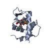


 PDBj
PDBj


 Assembly
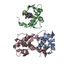

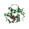
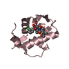

 Mass: 556.498 Da / Num. of mol.: 3 / Source method: obtained synthetically / Formula: C26H31Cl2NO6S
 Mass: 18.015 Da / Num. of mol.: 32 / Source method: isolated from a natural source / Formula: H2O
 Sample preparation
 / Beamline: 5.0.2 / Wavelength: 1 Å
 Processing