 Movie
Movie Controller

[English] 日本語
 Yorodumi
- PDB-4mul: Crystal structure of pantothenate synthetase in complex with 2-(5... -
+ Open data

- Basic information
| Entry | Database: PDB / ID: 4mul | ||||||
|---|---|---|---|---|---|---|---|
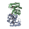
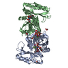
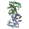
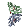
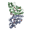
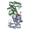
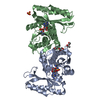
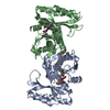
| Title | Crystal structure of pantothenate synthetase in complex with 2-(5-methoxy-2-(naphthalen-2-ylsulfonylcarbamoyl)-1H-indol-1-yl)acetic acid | ||||||
 Components Components | Pantothenate synthetase | ||||||
 Keywords Keywords | ligase/ligase inhibitor / Alpha Beta 3-Layer(aba) Sandwich Rossmann fold / Pantoate-ligase / ATP binding / ligase-ligase inhibitor complex | ||||||
| Function / homology |  Function and homology information Function and homology information: / pantoate-beta-alanine ligase (AMP-forming) / pantoate-beta-alanine ligase activity / pantothenate biosynthetic process / manganese ion binding / magnesium ion binding / ATP binding / metal ion binding / cytosol / cytoplasm Similarity search - Function | ||||||
| Biological species |   Mycobacterium tuberculosis (bacteria) Mycobacterium tuberculosis (bacteria) | ||||||
| Method |  X-RAY DIFFRACTION / SYNCHROTRON / MOLECULAR REPLACEMENT / Resolution: 1.75 Å X-RAY DIFFRACTION / SYNCHROTRON / MOLECULAR REPLACEMENT / Resolution: 1.75 Å | ||||||
 Authors Authors | Silvestre, H.L. / Blundell, T.L. | ||||||
 Citation Citation | Journal: Chemmedchem / Year: 2016 Title: Optimization of Inhibitors of Mycobacterium tuberculosis Pantothenate Synthetase Based on Group Efficiency Analysis. Authors: Hung, A.W. / Silvestre, H.L. / Wen, S. / George, G.P. / Boland, J. / Blundell, T.L. / Ciulli, A. / Abell, C. | ||||||
| History |
|
- Structure visualization
| Structure viewer | Molecule: MolmilJmol/JSmol |
|---|
- Downloads & links
-Download
| PDBx/mmCIF format | 4mul.cif.gz | 130.5 KB | Display | PDBx/mmCIF format |
|---|---|---|---|---|
| PDB format | pdb4mul.ent.gz | 101.7 KB | Display | PDB format |
| PDBx/mmJSON format | 4mul.json.gz | Tree view | PDBx/mmJSON format | |
| Others |  Other downloads Other downloads |
-Validation report
| Arichive directory | https://data.pdbj.org/pub/pdb/validation_reports/mu/4mulftp://data.pdbj.org/pub/pdb/validation_reports/mu/4mul | HTTPS FTP |
|---|
-Related structure data
| Related structure data |  4mq6C  4mueC  4mufC  4mugC  4muhC  4muiC  4mujC  4mukC  4munC  3le8S C: citing same article ( S: Starting model for refinement |
|---|---|
| Similar structure data |


-Links
 PDBj
PDBj


- Assembly
| Deposited unit | 
| ||||||||
|---|---|---|---|---|---|---|---|---|---|
| 1 |
| ||||||||
| 2 | 
| ||||||||
| Unit cell |
|
-Components
| #1: Protein | Mass: 31500.100 Da / Num. of mol.: 2 Source method: isolated from a genetically manipulated source Source: (gene. exp.) Mycobacterium tuberculosis (bacteria) / Gene: MT3707, MTCY07H7B.20, panC, Rv3602c / Plasmid: pET30b / Production host: References: UniProt: P0A5R0, UniProt: P9WIL5*PLUS, pantoate-beta-alanine ligase (AMP-forming) #2: Chemical | ChemComp-EOH /   Mass: 46.068 Da / Num. of mol.: 7 / Source method: obtained synthetically / Formula: C2H6O Mass: 46.068 Da / Num. of mol.: 7 / Source method: obtained synthetically / Formula: C2H6O#3: Chemical |   Mass: 438.453 Da / Num. of mol.: 3 / Source method: obtained synthetically / Formula: C22H18N2O6S Mass: 438.453 Da / Num. of mol.: 3 / Source method: obtained synthetically / Formula: C22H18N2O6S#4: Chemical | ChemComp-EDO /   Mass: 62.068 Da / Num. of mol.: 4 / Source method: obtained synthetically / Formula: C2H6O2 Mass: 62.068 Da / Num. of mol.: 4 / Source method: obtained synthetically / Formula: C2H6O2#5: Water | ChemComp-HOH / |  Mass: 18.015 Da / Num. of mol.: 411 / Source method: isolated from a natural source / Formula: H2O Mass: 18.015 Da / Num. of mol.: 411 / Source method: isolated from a natural source / Formula: H2O |
|---|
-Experimental details
-Experiment
| Experiment | Method: X-RAY DIFFRACTION / Number of used crystals: 1 |
|---|
- Sample preparation
| Crystal | Density Matthews: 2.21 Å3/Da / Density % sol: 44.39 % |
|---|---|
| Crystal grow | Temperature: 298 K / Method: vapor diffusion, hanging drop / pH: 8 Details: 11-14% w/v PEG3000, 150 mM lithium sulfate, 100 mM imidazole, 2% v/v ethanol, 10% v/v glycerol, 20 mM magnesium chloride, pH 8.0, VAPOR DIFFUSION, HANGING DROP, temperature 298K |
-Data collection
| Diffraction | Mean temperature: 100 K |
|---|---|
| Diffraction source | Source: SYNCHROTRON / Site: SLS  / Beamline: X06DA / Wavelength: 0.97 Å / Beamline: X06DA / Wavelength: 0.97 Å |
| Detector | Type: MARMOSAIC 325 mm CCD / Detector: CCD |
| Radiation | Protocol: SINGLE WAVELENGTH / Monochromatic (M) / Laue (L): M / Scattering type: x-ray |
| Radiation wavelength | Wavelength: 0.97 Å / Relative weight: 1 |
| Reflection | Resolution: 1.75→21.429 Å / Num. all: 204182 / Num. obs: 55392 / % possible obs: 99.9 % / Redundancy: 3.7 % / Biso Wilson estimate: 23 Å2 / Rmerge(I) obs: 0.062 / Net I/σ(I): 13.1 |
| Reflection shell | Resolution: 1.75→1.84 Å / Redundancy: 3.6 % / Rmerge(I) obs: 0.586 / Mean I/σ(I) obs: 2 / Num. unique all: 8058 / % possible all: 99.9 |
- Processing
| Software |
| ||||||||||||||||||||||||||||||||||||||||||||||||||||||||||||||||||||||||||||||||||||||||||||||||||||||||||||||||||||||||||||||||||||||||||||||||||||||||||||||||||||||||||||||||||||||
|---|---|---|---|---|---|---|---|---|---|---|---|---|---|---|---|---|---|---|---|---|---|---|---|---|---|---|---|---|---|---|---|---|---|---|---|---|---|---|---|---|---|---|---|---|---|---|---|---|---|---|---|---|---|---|---|---|---|---|---|---|---|---|---|---|---|---|---|---|---|---|---|---|---|---|---|---|---|---|---|---|---|---|---|---|---|---|---|---|---|---|---|---|---|---|---|---|---|---|---|---|---|---|---|---|---|---|---|---|---|---|---|---|---|---|---|---|---|---|---|---|---|---|---|---|---|---|---|---|---|---|---|---|---|---|---|---|---|---|---|---|---|---|---|---|---|---|---|---|---|---|---|---|---|---|---|---|---|---|---|---|---|---|---|---|---|---|---|---|---|---|---|---|---|---|---|---|---|---|---|---|---|---|---|
| Refinement | Method to determine structure: MOLECULAR REPLACEMENT Starting model: pdb entry 3LE8 Resolution: 1.75→21.429 Å / Cor.coef. Fo:Fc: 0.965 / Cor.coef. Fo:Fc free: 0.939 / SU B: 2.63 / SU ML: 0.084 / Cross valid method: THROUGHOUT / ESU R: 0.118 / ESU R Free: 0.122 / Stereochemistry target values: MAXIMUM LIKELIHOOD / Details: HYDROGENS HAVE BEEN ADDED IN THE RIDING POSITIONS
| ||||||||||||||||||||||||||||||||||||||||||||||||||||||||||||||||||||||||||||||||||||||||||||||||||||||||||||||||||||||||||||||||||||||||||||||||||||||||||||||||||||||||||||||||||||||
| Solvent computation | Ion probe radii: 0.8 Å / Shrinkage radii: 0.8 Å / VDW probe radii: 1.4 Å / Solvent model: MASK | ||||||||||||||||||||||||||||||||||||||||||||||||||||||||||||||||||||||||||||||||||||||||||||||||||||||||||||||||||||||||||||||||||||||||||||||||||||||||||||||||||||||||||||||||||||||
| Displacement parameters | Biso mean: 28.626 Å2
| ||||||||||||||||||||||||||||||||||||||||||||||||||||||||||||||||||||||||||||||||||||||||||||||||||||||||||||||||||||||||||||||||||||||||||||||||||||||||||||||||||||||||||||||||||||||
| Refinement step | Cycle: LAST / Resolution: 1.75→21.429 Å
| ||||||||||||||||||||||||||||||||||||||||||||||||||||||||||||||||||||||||||||||||||||||||||||||||||||||||||||||||||||||||||||||||||||||||||||||||||||||||||||||||||||||||||||||||||||||
| Refine LS restraints |
|