 Movie
Movie Controller

[English] 日本語
 Yorodumi
- PDB-4kmb: COMPLEX OF 4'-SULFO-LEWIS-X WITH A SELECTIN-LIKE MUTANT OF MANNOS... -
+ Open data

- Basic information
| Entry | Database: PDB / ID: 4kmb | |||||||||
|---|---|---|---|---|---|---|---|---|---|---|
| Title | COMPLEX OF 4'-SULFO-LEWIS-X WITH A SELECTIN-LIKE MUTANT OF MANNOSE-BINDING PROTEIN A | |||||||||
 Components Components | MANNOSE-BINDING PROTEIN-A | |||||||||
 Keywords Keywords | LECTIN | |||||||||
| Function / homology |  Function and homology information Function and homology informationcalcium-dependent carbohydrate binding / complement activation, lectin pathway / oligosaccharide binding / : / collagen trimer / surfactant homeostasis / phosphatidylinositol-4-phosphate binding / protein homotrimerization / D-mannose binding / polysaccharide binding ...calcium-dependent carbohydrate binding / complement activation, lectin pathway / oligosaccharide binding / : / collagen trimer / surfactant homeostasis / phosphatidylinositol-4-phosphate binding / protein homotrimerization / D-mannose binding / polysaccharide binding / complement activation, classical pathway / multivesicular body / positive regulation of phagocytosis / calcium-dependent protein binding / protease binding / defense response to Gram-positive bacterium / calcium ion binding / protein homodimerization activity / : / identical protein binding Similarity search - Function | |||||||||
| Biological species |  | |||||||||
| Method |  X-RAY DIFFRACTION / MOLECULAR REPLACEMENT / Resolution: 2 Å X-RAY DIFFRACTION / MOLECULAR REPLACEMENT / Resolution: 2 Å | |||||||||
 Authors Authors | Ng, K.K.-S. / Weis, W.I. | |||||||||
 Citation Citation | Journal: Biochemistry / Year: 1997 Title: Structure of a selectin-like mutant of mannose-binding protein complexed with sialylated and sulfated Lewis(x) oligosaccharides. Authors: Ng, K.K. / Weis, W.I. #1: Journal: J.Biol.Chem. / Year: 1996Title: Introduction of Selectin-Like Binding Specificity Into a Homologous Mannose-Binding Protein Authors: Blanck, O. / Iobst, S.T. / Gabel, C. / Drickamer, K. #2: Journal: Structure / Year: 1994Title: Trimeric Structure of a C-Type Mannose-Binding Protein Authors: Weis, W.I. / Drickamer, K. | |||||||||
| History |
|
- Structure visualization
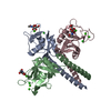
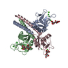
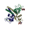
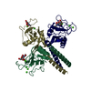
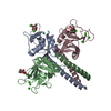
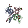
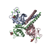
| Structure viewer | Molecule: MolmilJmol/JSmol |
|---|
- Downloads & links
-Download
| PDBx/mmCIF format | 4kmb.cif.gz | 117.3 KB | Display | PDBx/mmCIF format |
|---|---|---|---|---|
| PDB format | pdb4kmb.ent.gz | 88.3 KB | Display | PDB format |
| PDBx/mmJSON format | 4kmb.json.gz | Tree view | PDBx/mmJSON format | |
| Others |  Other downloads Other downloads |
-Validation report
| Arichive directory | https://data.pdbj.org/pub/pdb/validation_reports/km/4kmbftp://data.pdbj.org/pub/pdb/validation_reports/km/4kmb | HTTPS FTP |
|---|
-Related structure data
| Related structure data |  1kmbC 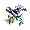 2kmbC  3kmbC  1rtmS S: Starting model for refinement C: citing same article ( |
|---|---|
| Similar structure data |


-Links
 PDBj
PDBj


- Assembly
| Deposited unit | 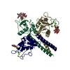
| ||||||||||||
|---|---|---|---|---|---|---|---|---|---|---|---|---|---|
| 1 |
| ||||||||||||
| Unit cell |
| ||||||||||||
| Noncrystallographic symmetry (NCS) | NCS oper:
|
-Components
-Protein , 1 types, 3 molecules 123
| #1: Protein | Mass: 16569.910 Da / Num. of mol.: 3 / Fragment: CLOSTRIPAIN FRAGMENT / Mutation: A211K, S212K, H213K Source method: isolated from a genetically manipulated source Source: (gene. exp.)  |
|---|
-Sugars , 2 types, 4 molecules 
| #2: Polysaccharide | Source method: isolated from a genetically manipulated source #6: Sugar |  Type: L-saccharide, alpha linking / Mass: 164.156 Da / Num. of mol.: 2 Type: L-saccharide, alpha linking / Mass: 164.156 Da / Num. of mol.: 2Source method: isolated from a genetically manipulated source Formula: C6H12O5 |
|---|
-Non-polymers , 4 types, 445 molecules 


| #3: Chemical | ChemComp-CA /  Mass: 40.078 Da / Num. of mol.: 11 / Source method: obtained synthetically / Formula: Ca Mass: 40.078 Da / Num. of mol.: 11 / Source method: obtained synthetically / Formula: Ca#4: Chemical |  Mass: 35.453 Da / Num. of mol.: 3 / Source method: obtained synthetically / Formula: Cl Mass: 35.453 Da / Num. of mol.: 3 / Source method: obtained synthetically / Formula: Cl#5: Chemical | ChemComp-ZN / |  Mass: 65.409 Da / Num. of mol.: 1 / Source method: obtained synthetically / Formula: Zn Mass: 65.409 Da / Num. of mol.: 1 / Source method: obtained synthetically / Formula: Zn#7: Water | ChemComp-HOH / | Mass: 18.015 Da / Num. of mol.: 430 / Source method: isolated from a natural source / Formula: H2O |
|---|
-Details
| Compound details | THE PURIFIED PROTEIN WAS DIGESTED WITH CLOSTRIPAIN TO PRODUCE THE FRAGMENT USED IN THE CRYSTAL ...THE PURIFIED PROTEIN WAS DIGESTED WITH CLOSTRIPAI |
|---|---|
| Has protein modification | Y |
-Experimental details
-Experiment
| Experiment | Method: X-RAY DIFFRACTION / Number of used crystals: 1 |
|---|
- Sample preparation
| Crystal | Density Matthews: 3.2 Å3/Da / Density % sol: 68 % | |||||||||||||||||||||||||||||||||||||||||||||||||||||||||||||||||||||||||||||
|---|---|---|---|---|---|---|---|---|---|---|---|---|---|---|---|---|---|---|---|---|---|---|---|---|---|---|---|---|---|---|---|---|---|---|---|---|---|---|---|---|---|---|---|---|---|---|---|---|---|---|---|---|---|---|---|---|---|---|---|---|---|---|---|---|---|---|---|---|---|---|---|---|---|---|---|---|---|---|
| Crystal grow | pH: 7.8 Details: PROTEIN WAS CRYSTALLIZED FROM 8-10% PEG 8000, 2% PEG 1000, 100 MM TRIS-CL, PH 7.8, 200 MM NACL, 20 MM CACL2, 2 MM NAN3. PRIOR TO DATA COLLECTION, THE CRYSTAL WAS ADAPTED TO THE MOTHER LIQUOR ...Details: PROTEIN WAS CRYSTALLIZED FROM 8-10% PEG 8000, 2% PEG 1000, 100 MM TRIS-CL, PH 7.8, 200 MM NACL, 20 MM CACL2, 2 MM NAN3. PRIOR TO DATA COLLECTION, THE CRYSTAL WAS ADAPTED TO THE MOTHER LIQUOR MINUS PEG 8000, PLUS 35% PEG 400, PLUS 80 MM 4'-SULFO- LEWIS-X. | |||||||||||||||||||||||||||||||||||||||||||||||||||||||||||||||||||||||||||||
| Crystal grow | *PLUS Temperature: 22 ℃ / Method: vapor diffusion | |||||||||||||||||||||||||||||||||||||||||||||||||||||||||||||||||||||||||||||
| Components of the solutions | *PLUS
|
-Data collection
| Diffraction | Mean temperature: 100 K |
|---|---|
| Diffraction source | Source: ROTATING ANODE / Type: RIGAKU RUH2R / Wavelength: 1.5418 |
| Detector | Type: RIGAKU RAXIS IIC / Detector: IMAGE PLATE / Date: May 28, 1996 / Details: COLLIMATOR |
| Radiation | Monochromator: GRAPHITE(002) / Monochromatic (M) / Laue (L): M / Scattering type: x-ray |
| Radiation wavelength | Wavelength: 1.5418 Å / Relative weight: 1 |
| Reflection | Resolution: 2→40 Å / Num. obs: 34603 / % possible obs: 97.5 % / Observed criterion σ(I): -3 / Redundancy: 2.2 % / Biso Wilson estimate: 20.6 Å2 / Rmerge(I) obs: 0.048 / Rsym value: 0.048 / Net I/σ(I): 12.2 |
| Reflection shell | Resolution: 2→2.07 Å / Redundancy: 2.1 % / Rmerge(I) obs: 0.242 / Mean I/σ(I) obs: 4.7 / Rsym value: 0.242 / % possible all: 94.7 |
| Reflection shell | *PLUS % possible obs: 94.7 % |
- Processing
| Software |
| ||||||||||||||||||||||||||||||||||||||||||||||||||||||||||||||||||||||||||||||||
|---|---|---|---|---|---|---|---|---|---|---|---|---|---|---|---|---|---|---|---|---|---|---|---|---|---|---|---|---|---|---|---|---|---|---|---|---|---|---|---|---|---|---|---|---|---|---|---|---|---|---|---|---|---|---|---|---|---|---|---|---|---|---|---|---|---|---|---|---|---|---|---|---|---|---|---|---|---|---|---|---|---|
| Refinement | Method to determine structure: MOLECULAR REPLACEMENT Starting model: 1RTM Resolution: 2→10 Å / Rfactor Rfree error: 0.004 / Data cutoff high absF: 100000 / Data cutoff low absF: 0.1 / Isotropic thermal model: RESTRAINED / Cross valid method: THROUGHOUT / σ(F): 2
| ||||||||||||||||||||||||||||||||||||||||||||||||||||||||||||||||||||||||||||||||
| Displacement parameters | Biso mean: 23.2 Å2
| ||||||||||||||||||||||||||||||||||||||||||||||||||||||||||||||||||||||||||||||||
| Refine analyze |
| ||||||||||||||||||||||||||||||||||||||||||||||||||||||||||||||||||||||||||||||||
| Refinement step | Cycle: LAST / Resolution: 2→10 Å
| ||||||||||||||||||||||||||||||||||||||||||||||||||||||||||||||||||||||||||||||||
| Refine LS restraints |
| ||||||||||||||||||||||||||||||||||||||||||||||||||||||||||||||||||||||||||||||||
| LS refinement shell | Resolution: 2→2.07 Å / Rfactor Rfree error: 0.017 / Total num. of bins used: 10
| ||||||||||||||||||||||||||||||||||||||||||||||||||||||||||||||||||||||||||||||||
| Xplor file |
| ||||||||||||||||||||||||||||||||||||||||||||||||||||||||||||||||||||||||||||||||
| Software | *PLUS Name: X-PLOR / Version: 3.54 / Classification: refinement | ||||||||||||||||||||||||||||||||||||||||||||||||||||||||||||||||||||||||||||||||
| Refine LS restraints | *PLUS
|
