 Movie
Movie Controller

[English] 日本語
 Yorodumi
- EMDB-4369: Unique features of mammalian mitochondrial translation initiation... -
+ Open data

- Basic information
| Entry | Database: EMDB / ID: EMD-4369 | ||||||||||||
|---|---|---|---|---|---|---|---|---|---|---|---|---|---|
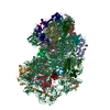
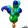
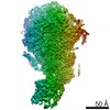
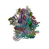
| Title | Unique features of mammalian mitochondrial translation initiation revealed by cryo-EM. This file contains the 28S ribosomal subunit. | ||||||||||||
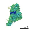
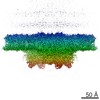
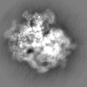
 Map data Map data | |||||||||||||
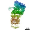
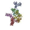
 Sample Sample |
| ||||||||||||
 Keywords Keywords | translation initiation / initiation factor IF2 / mitochondria / membrane targeting / RIBOSOME | ||||||||||||
| Function / homology |  Function and homology information Function and homology informationHormone ligand-binding receptors / gonadotropin hormone-releasing hormone activity / gonadotropin-releasing hormone receptor binding / G alpha (q) signalling events / Mitochondrial translation elongation / Mitochondrial ribosome-associated quality control / mitochondrial translational initiation / translation factor activity, RNA binding / formation of translation preinitiation complex / mitochondrial ribosome assembly ...Hormone ligand-binding receptors / gonadotropin hormone-releasing hormone activity / gonadotropin-releasing hormone receptor binding / G alpha (q) signalling events / Mitochondrial translation elongation / Mitochondrial ribosome-associated quality control / mitochondrial translational initiation / translation factor activity, RNA binding / formation of translation preinitiation complex / mitochondrial ribosome assembly / Mitochondrial translation termination / Mitochondrial protein degradation / Mitochondrial translation initiation / mitochondrial ribosome / ribosome disassembly / mitochondrial small ribosomal subunit / mitochondrial translation / organelle membrane / apoptotic mitochondrial changes / positive regulation of proteolysis / ribosomal small subunit binding / translation initiation factor activity / cell junction / regulation of translation / small ribosomal subunit / small ribosomal subunit rRNA binding / nuclear membrane / tRNA binding / mitochondrial inner membrane / rRNA binding / structural constituent of ribosome / ribosome / translation / mitochondrial matrix / ribonucleoprotein complex / mRNA binding / GTPase activity / GTP binding / nucleolus / mitochondrion / : / RNA binding / nucleoplasm / cytoplasm Similarity search - Function | ||||||||||||
| Biological species |   Homo sapiens (human) Homo sapiens (human) | ||||||||||||
| Method | single particle reconstruction / cryo EM / Resolution: 3.1 Å | ||||||||||||
 Authors Authors | Kummer E / Leibundgut M | ||||||||||||
| Funding support |  Switzerland, 3 items Switzerland, 3 items
| ||||||||||||
 Citation Citation | Journal: Nature / Year: 2018 Title: Unique features of mammalian mitochondrial translation initiation revealed by cryo-EM. Authors: Eva Kummer / Marc Leibundgut / Oliver Rackham / Richard G Lee / Daniel Boehringer / Aleksandra Filipovska / Nenad Ban /  Abstract: Mitochondria maintain their own specialized protein synthesis machinery, which in mammals is used exclusively for the synthesis of the membrane proteins responsible for oxidative phosphorylation. The ...Mitochondria maintain their own specialized protein synthesis machinery, which in mammals is used exclusively for the synthesis of the membrane proteins responsible for oxidative phosphorylation. The initiation of protein synthesis in mitochondria differs substantially from bacterial or cytosolic translation systems. Mitochondrial translation initiation lacks initiation factor 1, which is essential in all other translation systems from bacteria to mammals. Furthermore, only one type of methionyl transfer RNA (tRNA) is used for both initiation and elongation, necessitating that the initiation factor specifically recognizes the formylated version of tRNA (fMet-tRNA). Lastly, most mitochondrial mRNAs do not possess 5' leader sequences to promote mRNA binding to the ribosome. There is currently little mechanistic insight into mammalian mitochondrial translation initiation, and it is not clear how mRNA engagement, initiator-tRNA recruitment and start-codon selection occur. Here we determine the cryo-EM structure of the complete translation initiation complex from mammalian mitochondria at 3.2 Å. We describe the function of an additional domain insertion that is present in the mammalian mitochondrial initiation factor 2 (mtIF2). By closing the decoding centre, this insertion stabilizes the binding of leaderless mRNAs and induces conformational changes in the rRNA nucleotides involved in decoding. We identify unique features of mtIF2 that are required for specific recognition of fMet-tRNA and regulation of its GTPase activity. Finally, we observe that the ribosomal tunnel in the initiating ribosome is blocked by insertion of the N-terminal portion of mitochondrial protein mL45, which becomes exposed as the ribosome switches to elongation mode and may have an additional role in targeting of mitochondrial ribosomes to the protein-conducting pore in the inner mitochondrial membrane. | ||||||||||||
| History |
|
- Structure visualization
| Movie |
Movie viewer |
|---|---|
| Structure viewer | EM map: SurfViewMolmilJmol/JSmol |
| Supplemental images |

- Downloads & links
-EMDB archive
| Map data | emd_4369.map.gz | 12.7 MB | EMDB map data format | |
|---|---|---|---|---|
| Header (meta data) | emd-4369-v30.xmlemd-4369.xml | 66.1 KB 66.1 KB | Display Display | EMDB header |
| Images | 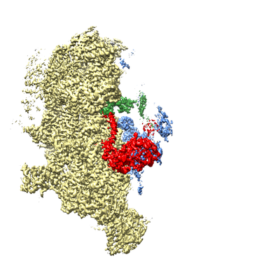 emd_4369.png emd_4369.png | 122.8 KB | ||
| Masks | emd_4369_msk_1.map | 84.6 MB | Mask map | |
| Filedesc metadata | emd-4369.cif.gz | 14.3 KB | ||
| Others | emd_4369_half_map_1.map.gzemd_4369_half_map_2.map.gz | 71.5 MB 71.5 MB | ||
| Archive directory |  http://ftp.pdbj.org/pub/emdb/structures/EMD-4369ftp://ftp.pdbj.org/pub/emdb/structures/EMD-4369 http://ftp.pdbj.org/pub/emdb/structures/EMD-4369ftp://ftp.pdbj.org/pub/emdb/structures/EMD-4369 | HTTPS FTP |
-Related structure data
| Related structure data |  6gazMC  4368C  4370C  6gawC 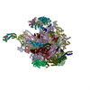 6gb2C C: citing same article ( M: atomic model generated by this map |
|---|---|
| Similar structure data |

-Links
| EMDB pages | EMDB (EBI/PDBe) / EMDataResource |
|---|---|
| Related items in Molecule of the Month |


-Map
| File | Download / File: emd_4369.map.gz / Format: CCP4 / Size: 84.6 MB / Type: IMAGE STORED AS FLOATING POINT NUMBER (4 BYTES) | ||||||||||||||||||||||||||||||||||||||||||||||||||||||||||||
|---|---|---|---|---|---|---|---|---|---|---|---|---|---|---|---|---|---|---|---|---|---|---|---|---|---|---|---|---|---|---|---|---|---|---|---|---|---|---|---|---|---|---|---|---|---|---|---|---|---|---|---|---|---|---|---|---|---|---|---|---|---|
| Projections & slices | Image control
Images are generated by Spider. | ||||||||||||||||||||||||||||||||||||||||||||||||||||||||||||
| Voxel size | X=Y=Z: 1.39 Å | ||||||||||||||||||||||||||||||||||||||||||||||||||||||||||||
| Density |
| ||||||||||||||||||||||||||||||||||||||||||||||||||||||||||||
| Symmetry | Space group: 1 | ||||||||||||||||||||||||||||||||||||||||||||||||||||||||||||
| Details | EMDB XML:
CCP4 map header:
| ||||||||||||||||||||||||||||||||||||||||||||||||||||||||||||
 Z (Sec.)
 Y (Row.)
 X (Col.)


-Supplemental data
-Mask #1
| File | emd_4369_msk_1.map | ||||||||||||
|---|---|---|---|---|---|---|---|---|---|---|---|---|---|
| Projections & Slices |
| ||||||||||||
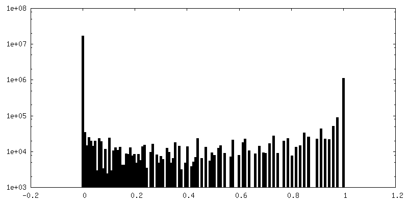
| Density Histograms |


-Half map: #1
| File | emd_4369_half_map_1.map | ||||||||||||
|---|---|---|---|---|---|---|---|---|---|---|---|---|---|
| Projections & Slices |
| ||||||||||||
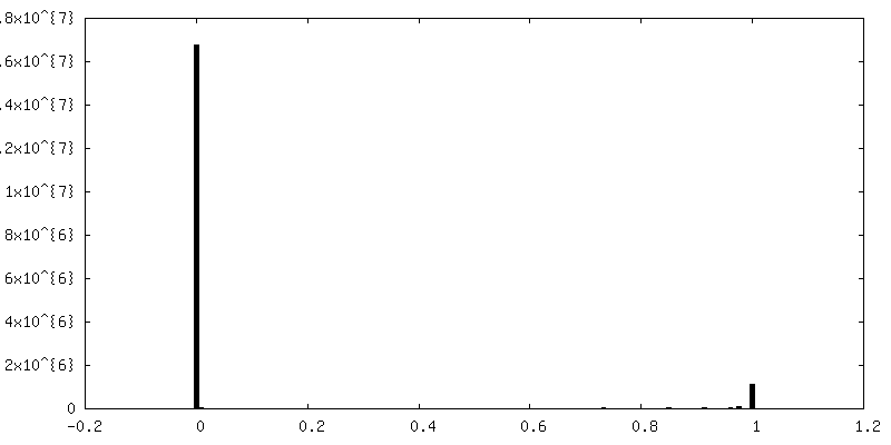
| Density Histograms |


-Half map: #2
| File | emd_4369_half_map_2.map | ||||||||||||
|---|---|---|---|---|---|---|---|---|---|---|---|---|---|
| Projections & Slices |
| ||||||||||||
| Density Histograms |


- Sample components
+Entire : mammalian mitochondrial translation initiation complex
+Supramolecule #1: mammalian mitochondrial translation initiation complex
+Supramolecule #2: Porcine Ribosome
+Supramolecule #3: Translation initiation factor IF-2, mitochondrial
+Supramolecule #4: Nucleic acids
+Macromolecule #1: Translation initiation factor IF-2, mitochondrial

+Macromolecule #2: Mitochondrial ribosomal protein L19
+Macromolecule #4: Mitochondrial ribosomal protein S2
+Macromolecule #5: Mitochondrial ribosomal protein S24
+Macromolecule #6: Mitochondrial ribosomal protein S5
+Macromolecule #7: Mitochondrial ribosomal protein S6
+Macromolecule #8: Mitochondrial ribosomal protein S7
+Macromolecule #9: Mitochondrial ribosomal protein S9
+Macromolecule #10: Mitochondrial ribosomal protein S10
+Macromolecule #11: Mitochondrial ribosomal protein S11
+Macromolecule #12: Mitochondrial ribosomal protein S12
+Macromolecule #13: Mitochondrial ribosomal protein S14
+Macromolecule #14: Mitochondrial ribosomal protein S15
+Macromolecule #15: bS16m, MRPS16
+Macromolecule #16: Mitochondrial ribosomal protein S17
+Macromolecule #17: Mitochondrial ribosomal protein S18C
+Macromolecule #18: Mitochondrial ribosomal protein S21
+Macromolecule #21: unassigned secondary structure elements
+Macromolecule #22: Mitochondrial ribosomal protein S22
+Macromolecule #23: Mitochondrial ribosomal protein S23
+Macromolecule #24: Mitochondrial ribosomal protein S25
+Macromolecule #25: Mitochondrial ribosomal protein S26
+Macromolecule #26: Mitochondrial ribosomal protein S27
+Macromolecule #27: Mitoribosomal protein ms28, mrps28
+Macromolecule #28: Death associated protein 3
+Macromolecule #29: mS31, MRPS31
+Macromolecule #30: Mitochondrial ribosomal protein S33
+Macromolecule #31: Mitochondrial ribosomal protein S34
+Macromolecule #32: Mitochondrial ribosomal protein S35
+Macromolecule #33: Mitochondrial ribosomal protein S37
+Macromolecule #34: Aurora kinase A interacting protein 1
+Macromolecule #35: Mitochondrial ribosomal protein S39
+Macromolecule #36: 28S ribosomal protein S18b, mitochondrial
+Macromolecule #3: 12S ribosomal RNA, mitochondrial
+Macromolecule #19: P-site fMet-tRNAMet, mitochondrial
+Macromolecule #20: MT-CO3 mRNA, mitochondrial
+Macromolecule #37: 5'-GUANOSINE-DIPHOSPHATE-MONOTHIOPHOSPHATE

+Macromolecule #38: MAGNESIUM ION
+Macromolecule #39: SODIUM ION
+Macromolecule #40: SPERMINE

+Macromolecule #41: ZINC ION
+Macromolecule #42: N-FORMYLMETHIONINE
+Macromolecule #43: GUANOSINE-5'-TRIPHOSPHATE

+Macromolecule #44: water

-Experimental details
-Structure determination
| Method | cryo EM |
|---|---|
 Processing Processing | single particle reconstruction |
| Aggregation state | particle |
-Sample preparation
| Concentration | 0.171 mg/mL |
|---|---|
| Buffer | pH: 7.6 |
| Grid | Model: Quantifoil R2/2 / Material: COPPER / Support film - Material: CARBON / Support film - topology: CONTINUOUS / Pretreatment - Type: GLOW DISCHARGE |
| Vitrification | Cryogen name: ETHANE / Chamber humidity: 100 % / Chamber temperature: 277.15 K / Instrument: FEI VITROBOT MARK IV |
| Details | contains 55S mitochondrial ribosome, mitochondrial initiation factor 2, mitochondrial formyl-Met-tRNAMet and MT-CO3 mRNA |
- Electron microscopy
| Microscope | FEI TITAN KRIOS |
|---|---|
| Image recording | Film or detector model: FEI FALCON III (4k x 4k) / Detector mode: INTEGRATING / Digitization - Dimensions - Width: 4096 pixel / Digitization - Dimensions - Height: 4096 pixel / Number real images: 13936 / Average exposure time: 1.4 sec. / Average electron dose: 40.0 e/Å2 |
| Electron beam | Acceleration voltage: 300 kV / Electron source:  FIELD EMISSION GUN FIELD EMISSION GUN |
| Electron optics | Illumination mode: FLOOD BEAM / Imaging mode: BRIGHT FIELD |
| Experimental equipment |  Model: Titan Krios / Image courtesy: FEI Company |
+Image processing
-Atomic model buiding 1
| Refinement | Space: RECIPROCAL / Protocol: OTHER / Overall B value: 73.8 |
|---|---|
| Output model | PDB-6gaz: |
