[English] 日本語
 Yorodumi
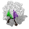
- EMDB-4126: Structure of the 70S ribosome with fMetSec-tRNASec in the hybrid ... -
+ Open data
- Basic information
| Entry | Database: EMDB / ID: EMD-4126 | |||||||||
|---|---|---|---|---|---|---|---|---|---|---|
| Title | Structure of the 70S ribosome with fMetSec-tRNASec in the hybrid pre-translocation state (H) | |||||||||
 Map data Map data | final sharpened map | |||||||||
 Sample Sample |
| |||||||||
 Keywords Keywords | translation / decoding / recoding / selenocysteine / ribosome | |||||||||
| Function / homology |  Function and homology information Function and homology informationnegative regulation of cytoplasmic translational initiation / stringent response / ornithine decarboxylase inhibitor activity / transcription antitermination factor activity, RNA binding / misfolded RNA binding / Group I intron splicing / RNA folding / transcriptional attenuation / endoribonuclease inhibitor activity / RNA-binding transcription regulator activity ...negative regulation of cytoplasmic translational initiation / stringent response / ornithine decarboxylase inhibitor activity / transcription antitermination factor activity, RNA binding / misfolded RNA binding / Group I intron splicing / RNA folding / transcriptional attenuation / endoribonuclease inhibitor activity / RNA-binding transcription regulator activity / positive regulation of ribosome biogenesis / negative regulation of cytoplasmic translation / translational termination / four-way junction DNA binding / DnaA-L2 complex / translation repressor activity / negative regulation of DNA-templated DNA replication initiation / negative regulation of translational initiation / regulation of mRNA stability / mRNA regulatory element binding translation repressor activity / ribosome assembly / assembly of large subunit precursor of preribosome / positive regulation of RNA splicing / transcription elongation factor complex / cytosolic ribosome assembly / regulation of DNA-templated transcription elongation / DNA endonuclease activity / response to reactive oxygen species / transcription antitermination / regulation of cell growth / translational initiation / DNA-templated transcription termination / maintenance of translational fidelity / response to radiation / mRNA 5'-UTR binding / ribosomal small subunit biogenesis / small ribosomal subunit rRNA binding / large ribosomal subunit / ribosome biogenesis / ribosome binding / regulation of translation / ribosomal small subunit assembly / small ribosomal subunit / 5S rRNA binding / large ribosomal subunit rRNA binding / transferase activity / cytosolic small ribosomal subunit / ribosomal large subunit assembly / cytoplasmic translation / cytosolic large ribosomal subunit / tRNA binding / molecular adaptor activity / negative regulation of translation / rRNA binding / ribosome / structural constituent of ribosome / translation / response to antibiotic / negative regulation of DNA-templated transcription / mRNA binding / DNA binding / RNA binding / zinc ion binding / membrane / cytosol / cytoplasm Similarity search - Function | |||||||||
| Biological species |  | |||||||||
| Method | single particle reconstruction / cryo EM / Resolution: 4.6 Å | |||||||||
 Authors Authors | Fischer N / Neumann P | |||||||||
| Funding support |  Germany, 2 items Germany, 2 items
| |||||||||
 Citation Citation |  Journal: Nature / Year: 2016 Journal: Nature / Year: 2016Title: The pathway to GTPase activation of elongation factor SelB on the ribosome. Authors: Niels Fischer / Piotr Neumann / Lars V Bock / Cristina Maracci / Zhe Wang / Alena Paleskava / Andrey L Konevega / Gunnar F Schröder / Helmut Grubmüller / Ralf Ficner / Marina V Rodnina / Holger Stark /  Abstract: In all domains of life, selenocysteine (Sec) is delivered to the ribosome by selenocysteine-specific tRNA (tRNA) with the help of a specialized translation factor, SelB in bacteria. Sec-tRNA recodes ...In all domains of life, selenocysteine (Sec) is delivered to the ribosome by selenocysteine-specific tRNA (tRNA) with the help of a specialized translation factor, SelB in bacteria. Sec-tRNA recodes a UGA stop codon next to a downstream mRNA stem-loop. Here we present the structures of six intermediates on the pathway of UGA recoding in Escherichia coli by single-particle cryo-electron microscopy. The structures explain the specificity of Sec-tRNA binding by SelB and show large-scale rearrangements of Sec-tRNA. Upon initial binding of SelB-Sec-tRNA to the ribosome and codon reading, the 30S subunit adopts an open conformation with Sec-tRNA covering the sarcin-ricin loop (SRL) on the 50S subunit. Subsequent codon recognition results in a local closure of the decoding site, which moves Sec-tRNA away from the SRL and triggers a global closure of the 30S subunit shoulder domain. As a consequence, SelB docks on the SRL, activating the GTPase of SelB. These results reveal how codon recognition triggers GTPase activation in translational GTPases. | |||||||||
| History |
|
- Structure visualization
| Movie |
 Movie viewer Movie viewer |
|---|---|
| Structure viewer | EM map:  SurfView SurfView Molmil Molmil Jmol/JSmol Jmol/JSmol |
| Supplemental images |
- Downloads & links
-EMDB archive
| Map data |  emd_4126.map.gz emd_4126.map.gz | 71.2 MB |  EMDB map data format EMDB map data format | |
|---|---|---|---|---|
| Header (meta data) |  emd-4126-v30.xml emd-4126-v30.xml emd-4126.xml emd-4126.xml | 73.6 KB 73.6 KB | Display Display |  EMDB header EMDB header |
| FSC (resolution estimation) |  emd_4126_fsc.xml emd_4126_fsc.xml | 9.4 KB | Display |  FSC data file FSC data file |
| Images |  emd_4126.png emd_4126.png | 148.1 KB | ||
| Filedesc metadata |  emd-4126.cif.gz emd-4126.cif.gz | 14.6 KB | ||
| Others |  emd_4126_half_map_1.map.gz emd_4126_half_map_1.map.gz emd_4126_half_map_2.map.gz emd_4126_half_map_2.map.gz | 60.1 MB 59.9 MB | ||
| Archive directory |  http://ftp.pdbj.org/pub/emdb/structures/EMD-4126 http://ftp.pdbj.org/pub/emdb/structures/EMD-4126 ftp://ftp.pdbj.org/pub/emdb/structures/EMD-4126 ftp://ftp.pdbj.org/pub/emdb/structures/EMD-4126 | HTTPS FTP |
-Validation report
| Summary document |  emd_4126_validation.pdf.gz emd_4126_validation.pdf.gz | 1.1 MB | Display |  EMDB validaton report EMDB validaton report |
|---|---|---|---|---|
| Full document |  emd_4126_full_validation.pdf.gz emd_4126_full_validation.pdf.gz | 1.1 MB | Display | |
| Data in XML |  emd_4126_validation.xml.gz emd_4126_validation.xml.gz | 17.1 KB | Display | |
| Data in CIF |  emd_4126_validation.cif.gz emd_4126_validation.cif.gz | 22.4 KB | Display | |
| Arichive directory |  https://ftp.pdbj.org/pub/emdb/validation_reports/EMD-4126 https://ftp.pdbj.org/pub/emdb/validation_reports/EMD-4126 ftp://ftp.pdbj.org/pub/emdb/validation_reports/EMD-4126 ftp://ftp.pdbj.org/pub/emdb/validation_reports/EMD-4126 | HTTPS FTP |
-Related structure data
| Related structure data |  5lzfMC  4121C  4122C  4123C  4124C  4125C  5lzaC  5lzbC  5lzcC  5lzdC  5lzeC M: atomic model generated by this map C: citing same article ( |
|---|---|
| Similar structure data | |
| EM raw data |  EMPIAR-10077 (Title: The pathway to GTPase activation of elongation factor SelB on the ribosome EMPIAR-10077 (Title: The pathway to GTPase activation of elongation factor SelB on the ribosomeData size: 1.0 TB Data #1: Part 1 - Unprocessed cryo-EM micrographs of E. Coli 70S-SelB-GDPNP-Sec-tRNASec-fMet-tRNAfMet-SECIS mRNA complexes [micrographs - single frame] Data #2: Part 2 - Unprocessed cryo-EM micrographs of E. Coli 70S-SelB-GDPNP-Sec-tRNASec-fMet-tRNAfMet-SECIS mRNA complex [micrographs - single frame] Data #3: ribosomeSelB_particles.mrcs - CTF corrected single particle images of E. Coli 70S-SelB-GDPNP-Sec-tRNASec-fMet-tRNAfMet-SECIS mRNA complex [picked particles - multiframe - processed]) |
- Links
Links
| EMDB pages |  EMDB (EBI/PDBe) / EMDB (EBI/PDBe) /  EMDataResource EMDataResource |
|---|---|
| Related items in Molecule of the Month |
- Map
Map
| File |  Download / File: emd_4126.map.gz / Format: CCP4 / Size: 76.8 MB / Type: IMAGE STORED AS FLOATING POINT NUMBER (4 BYTES) Download / File: emd_4126.map.gz / Format: CCP4 / Size: 76.8 MB / Type: IMAGE STORED AS FLOATING POINT NUMBER (4 BYTES) | ||||||||||||||||||||||||||||||||||||||||||||||||||||||||||||||||||||
|---|---|---|---|---|---|---|---|---|---|---|---|---|---|---|---|---|---|---|---|---|---|---|---|---|---|---|---|---|---|---|---|---|---|---|---|---|---|---|---|---|---|---|---|---|---|---|---|---|---|---|---|---|---|---|---|---|---|---|---|---|---|---|---|---|---|---|---|---|---|
| Annotation | final sharpened map | ||||||||||||||||||||||||||||||||||||||||||||||||||||||||||||||||||||
| Projections & slices | Image control
Images are generated by Spider. | ||||||||||||||||||||||||||||||||||||||||||||||||||||||||||||||||||||
| Voxel size | X=Y=Z: 1.16 Å | ||||||||||||||||||||||||||||||||||||||||||||||||||||||||||||||||||||
| Density |
| ||||||||||||||||||||||||||||||||||||||||||||||||||||||||||||||||||||
| Symmetry | Space group: 1 | ||||||||||||||||||||||||||||||||||||||||||||||||||||||||||||||||||||
| Details | EMDB XML:
CCP4 map header:
| ||||||||||||||||||||||||||||||||||||||||||||||||||||||||||||||||||||
-Supplemental data
-Half map: unfiltered half-map 1 (unprocessed)
| File | emd_4126_half_map_1.map | ||||||||||||
|---|---|---|---|---|---|---|---|---|---|---|---|---|---|
| Annotation | unfiltered half-map 1 (unprocessed) | ||||||||||||
| Projections & Slices |
| ||||||||||||
| Density Histograms |
-Half map: unfiltered half-map 2 (unprocessed)
| File | emd_4126_half_map_2.map | ||||||||||||
|---|---|---|---|---|---|---|---|---|---|---|---|---|---|
| Annotation | unfiltered half-map 2 (unprocessed) | ||||||||||||
| Projections & Slices |
| ||||||||||||
| Density Histograms |
- Sample components
+Entire : Hybrid pre-translocation state of E. coli ribosome 70S-fMetSec-tR...
+Supramolecule #1: Hybrid pre-translocation state of E. coli ribosome 70S-fMetSec-tR...
+Macromolecule #1: 16S ribosomal RNA
+Macromolecule #22: tRNAfMet
+Macromolecule #23: SECIS mRNA
+Macromolecule #24: fMetSec-tRNASec
+Macromolecule #25: 23S ribosomal RNA
+Macromolecule #26: 5S ribosomal RNA
+Macromolecule #2: 30S ribosomal protein S2
+Macromolecule #3: 30S ribosomal protein S3
+Macromolecule #4: 30S ribosomal protein S4
+Macromolecule #5: 30S ribosomal protein S5
+Macromolecule #6: 30S ribosomal protein S6
+Macromolecule #7: 30S ribosomal protein S7
+Macromolecule #8: 30S ribosomal protein S8
+Macromolecule #9: 30S ribosomal protein S9
+Macromolecule #10: 30S ribosomal protein S10
+Macromolecule #11: 30S ribosomal protein S11
+Macromolecule #12: 30S ribosomal protein S12
+Macromolecule #13: 30S ribosomal protein S13
+Macromolecule #14: 30S ribosomal protein S14
+Macromolecule #15: 30S ribosomal protein S15
+Macromolecule #16: 30S ribosomal protein S16
+Macromolecule #17: 30S ribosomal protein S17
+Macromolecule #18: 30S ribosomal protein S18
+Macromolecule #19: 30S ribosomal protein S19
+Macromolecule #20: 30S ribosomal protein S20
+Macromolecule #21: 30S ribosomal protein S21
+Macromolecule #27: 50S ribosomal protein L2
+Macromolecule #28: 50S ribosomal protein L3
+Macromolecule #29: 50S ribosomal protein L4
+Macromolecule #30: 50S ribosomal protein L5
+Macromolecule #31: 50S ribosomal protein L6
+Macromolecule #32: 50S ribosomal protein L11
+Macromolecule #33: 50S ribosomal protein L9
+Macromolecule #34: 50S ribosomal protein L13
+Macromolecule #35: 50S ribosomal protein L14
+Macromolecule #36: 50S ribosomal protein L15
+Macromolecule #37: 50S ribosomal protein L16
+Macromolecule #38: 50S ribosomal protein L17
+Macromolecule #39: 50S ribosomal protein L18
+Macromolecule #40: 50S ribosomal protein L19
+Macromolecule #41: 50S ribosomal protein L20
+Macromolecule #42: 50S ribosomal protein L21
+Macromolecule #43: 50S ribosomal protein L22
+Macromolecule #44: 50S ribosomal protein L23
+Macromolecule #45: 50S ribosomal protein L24
+Macromolecule #46: 50S ribosomal protein L25
+Macromolecule #47: 50S ribosomal protein L27
+Macromolecule #48: 50S ribosomal protein L28
+Macromolecule #49: 50S ribosomal protein L29
+Macromolecule #50: 50S ribosomal protein L30
+Macromolecule #51: 50S ribosomal protein L32
+Macromolecule #52: 50S ribosomal protein L33
+Macromolecule #53: 50S ribosomal protein L34
+Macromolecule #54: 50S ribosomal protein L35
+Macromolecule #55: 50S ribosomal protein L36
+Macromolecule #56: 50S ribosomal protein L31
+Macromolecule #57: ZINC ION
-Experimental details
-Structure determination
| Method | cryo EM |
|---|---|
 Processing Processing | single particle reconstruction |
| Aggregation state | particle |
- Sample preparation
| Buffer | pH: 7.5 Details: 50 mM Hepes-KOH, pH 7.5, 70 mM NH4Cl, 30 mM KCl, 7 mM MgCl2, 0.6mM spermine, 0.4mM spermidine, 2 mM DTT |
|---|---|
| Grid | Model: Quantifoil R3.5/1 / Material: COPPER / Support film - Material: CARBON / Support film - topology: CONTINUOUS / Pretreatment - Type: GLOW DISCHARGE |
| Vitrification | Cryogen name: ETHANE / Chamber humidity: 100 % / Instrument: FEI VITROBOT MARK IV |
- Electron microscopy
| Microscope | FEI TITAN KRIOS |
|---|---|
| Specialist optics | Spherical aberration corrector: CEOS Cs-corrector |
| Details | Using a Cs-corrector from CEOS electron optical aberrations were corrected to residual phase errors of 45degree at scattering angles of >12 to 15 mrad. |
| Image recording | Film or detector model: FEI FALCON II (4k x 4k) / Detector mode: INTEGRATING / Number grids imaged: 1 / Number real images: 12681 / Average exposure time: 1.0 sec. / Average electron dose: 30.0 e/Å2 |
| Electron beam | Acceleration voltage: 300 kV / Electron source:  FIELD EMISSION GUN FIELD EMISSION GUN |
| Electron optics | Illumination mode: SPOT SCAN / Imaging mode: BRIGHT FIELD / Cs: 0.001 mm / Nominal defocus max: 2.6 µm / Nominal defocus min: 0.7000000000000001 µm / Nominal magnification: 59000 |
| Sample stage | Specimen holder model: FEI TITAN KRIOS AUTOGRID HOLDER / Cooling holder cryogen: NITROGEN |
| Experimental equipment |  Model: Titan Krios / Image courtesy: FEI Company |
+ Image processing
-Atomic model buiding 1
| Details | For parts of the model exhibiting larger conformational differences and/or lower local map resolution, additional cycles of real space refinement and manual fitting were performed against experimental map filtered to lower resolution |
|---|---|
| Refinement | Protocol: FLEXIBLE FIT / Target criteria: Maximum likelihood |
| Output model |  PDB-5lzf: |
 Movie
Movie Controller


 Z (Sec.)
 Y (Row.)
 X (Col.)


