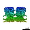
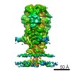
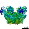
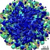
ジャーナル: PLoS Pathog / 年: 2016 タイトル: Cryo-electron Microscopy Structure of the Native Prototype Foamy Virus Glycoprotein and Virus Architecture. 著者: Grégory Effantin / Leandro F Estrozi / Nick Aschman / Patricia Renesto / Nicole Stanke / Dirk Lindemann / Guy Schoehn / Winfried Weissenhorn / 要旨: Foamy viruses (FV) belong to the genus Spumavirus, which forms a distinct lineage in the Retroviridae family. Although the infection in natural hosts and zoonotic transmission to humans is ...Foamy viruses (FV) belong to the genus Spumavirus, which forms a distinct lineage in the Retroviridae family. Although the infection in natural hosts and zoonotic transmission to humans is asymptomatic, FVs can replicate well in human cells making it an attractive gene therapy vector candidate. Here we present cryo-electron microscopy and (cryo-)electron tomography ultrastructural data on purified prototype FV (PFV) and PFV infected cells. Mature PFV particles have a distinct morphology with a capsid of constant dimension as well as a less ordered shell of density between the capsid and the membrane likely formed by the Gag N-terminal domain and the cytoplasmic part of the Env leader peptide gp18LP. The viral membrane contains trimeric Env glycoproteins partly arranged in interlocked hexagonal assemblies. In situ 3D reconstruction by subtomogram averaging of wild type Env and of a Env gp48TM- gp80SU cleavage site mutant showed a similar spike architecture as well as stabilization of the hexagonal lattice by clear connections between lower densities of neighboring trimers. Cryo-EM was employed to obtain a 9 Å resolution map of the glycoprotein in its pre-fusion state, which revealed extensive trimer interactions by the receptor binding subunit gp80SU at the top of the spike and three central helices derived from the fusion protein subunit gp48TM. The lower part of Env, presumably composed of interlaced parts of gp48TM, gp80SU and gp18LP anchors the spike at the membrane. We propose that the gp48TM density continues into three central transmembrane helices, which interact with three outer transmembrane helices derived from gp18LP. Our ultrastructural data and 9 Å resolution glycoprotein structure provide important new insights into the molecular architecture of PFV and its distinct evolutionary relationship with other members of the Retroviridae.
名称: Human spumaretrovirus / タイプ: virus / ID: 1 / 親要素: 0 / 詳細: Mutant in the Gag polyprotein / NCBI-ID: 11963 / 生物種: Human spumaretrovirus / ウイルスタイプ: VIRION / ウイルス・単離状態: SPECIES / ウイルス・エンベロープ: Yes / ウイルス・中空状態: No
Host system
生物種: Homo sapiens (ヒト) / 組換プラスミド: pcoPG4 GR R/A, pcoPE32, pcoPP
分子量
理論値: 330 KDa
-
実験情報
-
構造解析
手法
クライオ電子顕微鏡法
解析
単粒子再構成法
試料の集合状態
particle
-
試料調製
緩衝液
pH: 7.5
凍結
凍結剤: ETHANE
詳細
In the iNAB mutant,23 arginines in the glycine/arginine rich (GR) region in the C-terminus of Gag have been replaced by alanine, which results in a Gag protein unable to bind nucleic acid. Virus particles are still released from cells, although less efficiently, but are non-infectious and display capsid assembly defects.
-
電子顕微鏡法
顕微鏡
FEI TECNAI F30
撮影
フィルム・検出器のモデル: GATAN K2 SUMMIT (4k x 4k) 平均電子線量: 30.0 e/Å2
電子線
加速電圧: 300 kV / 電子線源: FIELD EMISSION GUN
電子光学系
照射モード: FLOOD BEAM / 撮影モード: BRIGHT FIELD
実験機器
モデル: Tecnai F30 / 画像提供: FEI Company
+
画像解析
CTF補正
ソフトウェア - 名称: CTFFIND (ver. 4)
初期モデル
モデルのタイプ: OTHER / 詳細: Initial model is from subtomogram averaging
 ムービー
ムービー コントローラー


 データを開く
 基本情報
基本情報 マップデータ
 試料
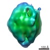
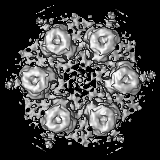
試料 Human spumaretrovirus (ウイルス)
 データ登録者
 引用
引用

 構造の表示
構造の表示 ムービービューア
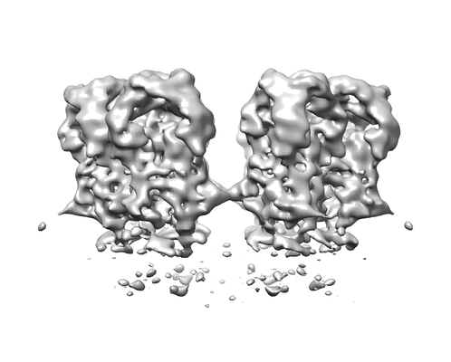
 ダウンロードとリンク
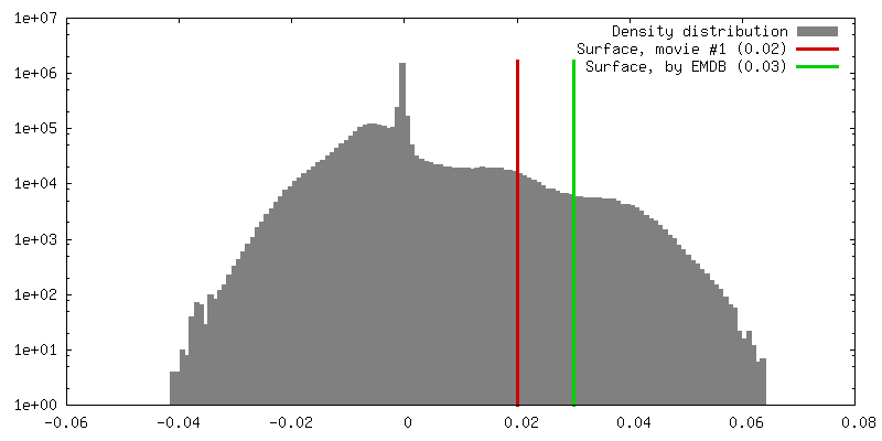
 emd_4010.png
 http://ftp.pdbj.org/pub/emdb/structures/EMD-4010


 Z (Sec.)
 Y (Row.)
 X (Col.)


 試料の構成要素
 Homo sapiens (ヒト) / 組換プラスミド: pcoPG4 GR R/A, pcoPE32, pcoPP
 解析
解析 電子顕微鏡法
 FIELD EMISSION GUN
