 Movie
Movie Controller


+ Open data

- Basic information
| Entry | Database: PDB / ID: 3jc1 | ||||||
|---|---|---|---|---|---|---|---|
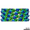
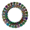
| Title | Electron cryo-microscopy of the IST1-CHMP1B ESCRT-III copolymer | ||||||
 Components Components |
| ||||||
 Keywords Keywords | LIPID BINDING PROTEIN / ESCRT-III / IST1 / CHMP1B / membrane tubulation / helical filament | ||||||
| Function / homology |  Function and homology information Function and homology informationMIT domain binding / amphisome membrane / multivesicular body-lysosome fusion / vesicle fusion with vacuole / abscission / ESCRT III complex disassembly / late endosome to lysosome transport / ESCRT III complex / kinetochore microtubule / endosome transport via multivesicular body sorting pathway ...MIT domain binding / amphisome membrane / multivesicular body-lysosome fusion / vesicle fusion with vacuole / abscission / ESCRT III complex disassembly / late endosome to lysosome transport / ESCRT III complex / kinetochore microtubule / endosome transport via multivesicular body sorting pathway / cytoskeleton-dependent cytokinesis / regulation of centrosome duplication / nuclear membrane reassembly / collateral sprouting / Sealing of the nuclear envelope (NE) by ESCRT-III / midbody abscission / positive regulation of collateral sprouting / multivesicular body sorting pathway / membrane fission / plasma membrane repair / membrane coat / late endosome to vacuole transport / multivesicular body membrane / ubiquitin-dependent protein catabolic process via the multivesicular body sorting pathway / regulation of mitotic spindle assembly / multivesicular body assembly / Flemming body / mitotic metaphase chromosome alignment / nucleus organization / viral budding via host ESCRT complex / positive regulation of proteolysis / autophagosome membrane / endoplasmic reticulum-Golgi intermediate compartment / autophagosome maturation / nuclear pore / multivesicular body / viral budding from plasma membrane / establishment of protein localization / kinetochore / autophagy / azurophil granule lumen / protein localization / protein transport / nuclear envelope / midbody / endosome membrane / cadherin binding / protein domain specific binding / lysosomal membrane / cell division / intracellular membrane-bounded organelle / centrosome / Neutrophil degranulation / protein-containing complex binding / chromatin / extracellular exosome / extracellular region / nucleoplasm / identical protein binding / plasma membrane / cytosol Similarity search - Function | ||||||
| Biological species |  Homo sapiens (human) Homo sapiens (human) | ||||||
| Method | ELECTRON MICROSCOPY / helical reconstruction / cryo EM / Resolution: 4 Å | ||||||
 Authors Authors | McCullough, J. / Clippinger, A.K. / Talledge, N. / Skowyra, M.L. / Saunders, M.G. / Naismith, T.V. / Colf, L.A. / Afonine, P. / Arthur, C. / Sundquist, W.I. ...McCullough, J. / Clippinger, A.K. / Talledge, N. / Skowyra, M.L. / Saunders, M.G. / Naismith, T.V. / Colf, L.A. / Afonine, P. / Arthur, C. / Sundquist, W.I. / Hanson, P.I. / Frost, A. | ||||||
 Citation Citation | Journal: Science / Year: 2015 Title: Structure and membrane remodeling activity of ESCRT-III helical polymers. Authors: John McCullough / Amy K Clippinger / Nathaniel Talledge / Michael L Skowyra / Marissa G Saunders / Teresa V Naismith / Leremy A Colf / Pavel Afonine / Christopher Arthur / Wesley I Sundquist ...Authors: John McCullough / Amy K Clippinger / Nathaniel Talledge / Michael L Skowyra / Marissa G Saunders / Teresa V Naismith / Leremy A Colf / Pavel Afonine / Christopher Arthur / Wesley I Sundquist / Phyllis I Hanson / Adam Frost /  Abstract: The endosomal sorting complexes required for transport (ESCRT) proteins mediate fundamental membrane remodeling events that require stabilizing negative membrane curvature. These include endosomal ...The endosomal sorting complexes required for transport (ESCRT) proteins mediate fundamental membrane remodeling events that require stabilizing negative membrane curvature. These include endosomal intralumenal vesicle formation, HIV budding, nuclear envelope closure, and cytokinetic abscission. ESCRT-III subunits perform key roles in these processes by changing conformation and polymerizing into membrane-remodeling filaments. Here, we report the 4 angstrom resolution cryogenic electron microscopy reconstruction of a one-start, double-stranded helical copolymer composed of two different human ESCRT-III subunits, charged multivesicular body protein 1B (CHMP1B) and increased sodium tolerance 1 (IST1). The inner strand comprises "open" CHMP1B subunits that interlock in an elaborate domain-swapped architecture and is encircled by an outer strand of "closed" IST1 subunits. Unlike other ESCRT-III proteins, CHMP1B and IST1 polymers form external coats on positively curved membranes in vitro and in vivo. Our analysis suggests how common ESCRT-III filament architectures could stabilize different degrees and directions of membrane curvature. | ||||||
| History |
|
- Structure visualization
| Movie |
Movie viewer |
|---|---|
| Structure viewer | Molecule: MolmilJmol/JSmol |
 UCSF Chimera
- Downloads & links
-Download
| PDBx/mmCIF format | 3jc1.cif.gz | 1.8 MB | Display | PDBx/mmCIF format |
|---|---|---|---|---|
| PDB format | pdb3jc1.ent.gz | Display | PDB format | |
| PDBx/mmJSON format | 3jc1.json.gz | Tree view | PDBx/mmJSON format | |
| Others |  Other downloads Other downloads |
-Validation report
| Summary document | 3jc1_validation.pdf.gz | 1.6 MB | Display | wwPDB validaton report |
|---|---|---|---|---|
| Full document | 3jc1_full_validation.pdf.gz | 1.6 MB | Display | |
| Data in XML | 3jc1_validation.xml.gz | 241.2 KB | Display | |
| Data in CIF | 3jc1_validation.cif.gz | 358 KB | Display | |
| Arichive directory | https://data.pdbj.org/pub/pdb/validation_reports/jc/3jc1ftp://data.pdbj.org/pub/pdb/validation_reports/jc/3jc1 | HTTPS FTP |
-Related structure data
| Related structure data | 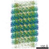 6461MC M: map data used to model this data C: citing same article ( |
|---|---|
| Similar structure data |


-Links
 PDBj
PDBj- Assembly
| Deposited unit | 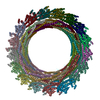
|
|---|---|
| 1 |
|
| Symmetry | Helical symmetry: (Circular symmetry: 1 / Dyad axis: no / N subunits divisor: 1 / Num. of operations: 34 / Rise per n subunits: 2.96 Å / Rotation per n subunits: 21.06 °) |
-Components
| #1: Protein | Mass: 20934.514 Da / Num. of mol.: 34 / Fragment: N-terminal domain (UNP residues 6-187) Source method: isolated from a genetically manipulated source Source: (gene. exp.) Homo sapiens (human) / Gene: IST1, KIAA0174 / Plasmid: PGEX-2T-TEV / Production host:  #2: Protein | Mass: 17937.719 Da / Num. of mol.: 34 / Fragment: UNP residues 4-163 Source method: isolated from a genetically manipulated source Source: (gene. exp.) Homo sapiens (human) / Gene: C18orf2, CHMP1B / Plasmid: PGEX-2T-TEV / Production host: |
|---|
-Experimental details
-Experiment
| Experiment | Method: ELECTRON MICROSCOPY |
|---|---|
| EM experiment | Aggregation state: FILAMENT / 3D reconstruction method: helical reconstruction |
- Sample preparation
| Component |
| ||||||||||||||||
|---|---|---|---|---|---|---|---|---|---|---|---|---|---|---|---|---|---|
| Buffer solution | Name: 25 mM Tris, pH 8.0, 25 mM sodium chloride / pH: 8 / Details: 25 mM Tris, pH 8.0, 25 mM sodium chloride | ||||||||||||||||
| Specimen | Embedding applied: NO / Shadowing applied: NO / Staining applied: NO / Vitrification applied: YES | ||||||||||||||||
| Specimen support | Details: Quantifoil R2/2 200 Mesh (glow-discharged) holey carbon grids | ||||||||||||||||
| Vitrification | Instrument: FEI VITROBOT MARK III / Cryogen name: ETHANE / Humidity: 100 % / Chamber temperature: 298 K Details: Deposited 3.5 uL sample, blotted 3-6 seconds (0 mm offset), and plunged into liquid ethane (VITROBOT MARK III). Method: Deposited 3.5 uL sample, blotted 3-6 seconds (0 mm offset) |
- Electron microscopy imaging
| Experimental equipment |  Model: Titan Krios / Image courtesy: FEI Company /  Model: Tecnai F20 / Image courtesy: FEI Company | ||||||||||||||||||||||||
|---|---|---|---|---|---|---|---|---|---|---|---|---|---|---|---|---|---|---|---|---|---|---|---|---|---|
| EM imaging | Date: Jun 1, 2013 / Electron source:
| ||||||||||||||||||||||||
| Image recording | Electron dose: 20 e/Å2 / Film or detector model: KODAK SO-163 FILM |
- Processing
| EM software |
| ||||||||||||||||||||||||||||||||||||||||||||
|---|---|---|---|---|---|---|---|---|---|---|---|---|---|---|---|---|---|---|---|---|---|---|---|---|---|---|---|---|---|---|---|---|---|---|---|---|---|---|---|---|---|---|---|---|---|
| CTF correction | Details: CTFFIND3 | ||||||||||||||||||||||||||||||||||||||||||||
| Helical symmerty | Angular rotation/subunit: 21.06 ° / Axial rise/subunit: 2.96 Å / Axial symmetry: C1 | ||||||||||||||||||||||||||||||||||||||||||||
| 3D reconstruction | Method: Iterative Helical Real Space Reconstruction (IHRSR) / Resolution: 4 Å / Num. of particles: 188713 / Nominal pixel size: 1.22 Å / Actual pixel size: 1.22 Å Details: Modified version of IHRSR algorithms as implemented in SPIDER was used to determine the helical symmetry. 3D reconstructions were performed using RELION. Symmetry type: HELICAL | ||||||||||||||||||||||||||||||||||||||||||||
| Atomic model building | Space: REAL / Target criteria: Molprobity validation, cross-correlation Details: DETAILS--IST1 starting model (3FRR) was idealized in Rosetta, then relaxed while the structure was constrained. Iterative loop building was used. | ||||||||||||||||||||||||||||||||||||||||||||
| Atomic model building | PDB-ID: 3FRR Accession code: 3FRR / Source name: PDB / Type: experimental model | ||||||||||||||||||||||||||||||||||||||||||||
| Refinement step | Cycle: LAST
|

