 Movie
Movie Controller

[English] 日本語
 Yorodumi
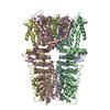
- PDB-3j9p: Structure of the TRPA1 ion channel determined by electron cryo-mi... -
+ Open data

- Basic information
| Entry | Database: PDB / ID: 3j9p | ||||||
|---|---|---|---|---|---|---|---|
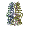
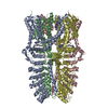
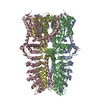
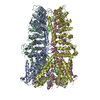
| Title | Structure of the TRPA1 ion channel determined by electron cryo-microscopy | ||||||
 Components Components | Maltose-binding periplasmic protein, Transient receptor potential cation channel subfamily A member 1 chimera | ||||||
 Keywords Keywords | TRANSPORT PROTEIN / TRPA1 / TRP / transient / potential / receptor / ion channel / membrane protein | ||||||
| Function / homology |  Function and homology information Function and homology informationregulation of blood circulation / positive regulation of monoatomic anion transport / cellular response to food / temperature-gated cation channel activity / regulation of neuronal action potential / cellular response to carbon dioxide / osmolarity-sensing monoatomic cation channel activity / detection of mechanical stimulus involved in sensory perception / stereocilium bundle / detection of chemical stimulus involved in sensory perception of pain ...regulation of blood circulation / positive regulation of monoatomic anion transport / cellular response to food / temperature-gated cation channel activity / regulation of neuronal action potential / cellular response to carbon dioxide / osmolarity-sensing monoatomic cation channel activity / detection of mechanical stimulus involved in sensory perception / stereocilium bundle / detection of chemical stimulus involved in sensory perception of pain / urinary bladder smooth muscle contraction / thermoception / cellular response to toxic substance / cellular response to caffeine / cellular response to cold / calcium ion transmembrane import into cytosol / detection of maltose stimulus / TRP channels / maltose transport complex / intracellularly gated calcium channel activity / carbohydrate transport / carbohydrate transmembrane transporter activity / maltose binding / maltose transport / maltodextrin transmembrane transport / monoatomic ion transport / voltage-gated calcium channel activity / ATP-binding cassette (ABC) transporter complex, substrate-binding subunit-containing / response to cold / ATP-binding cassette (ABC) transporter complex / sensory perception of pain / positive regulation of insulin secretion involved in cellular response to glucose stimulus / cell chemotaxis / intracellular calcium ion homeostasis / cellular response to hydrogen peroxide / calcium ion transmembrane transport / calcium channel activity / cellular response to heat / outer membrane-bounded periplasmic space / channel activity / protein homotetramerization / periplasmic space / cell surface receptor signaling pathway / apical plasma membrane / axon / DNA damage response / membrane / identical protein binding / plasma membrane Similarity search - Function | ||||||
| Biological species |   Homo sapiens (human) Homo sapiens (human) | ||||||
| Method | ELECTRON MICROSCOPY / single particle reconstruction / cryo EM / Resolution: 4.24 Å | ||||||
 Authors Authors | Paulsen, C.E. / Armache, J.-P. / Gao, Y. / Cheng, Y. / Julius, D. | ||||||
 Citation Citation | Journal: Nature / Year: 2015 Title: Structure of the TRPA1 ion channel suggests regulatory mechanisms. Authors: Candice E Paulsen / Jean-Paul Armache / Yuan Gao / Yifan Cheng / David Julius /  Abstract: The TRPA1 ion channel (also known as the wasabi receptor) is a detector of noxious chemical agents encountered in our environment or produced endogenously during tissue injury or drug metabolism. ...The TRPA1 ion channel (also known as the wasabi receptor) is a detector of noxious chemical agents encountered in our environment or produced endogenously during tissue injury or drug metabolism. These include a broad class of electrophiles that activate the channel through covalent protein modification. TRPA1 antagonists hold potential for treating neurogenic inflammatory conditions provoked or exacerbated by irritant exposure. Despite compelling reasons to understand TRPA1 function, structural mechanisms underlying channel regulation remain obscure. Here we use single-particle electron cryo- microscopy to determine the structure of full-length human TRPA1 to ∼4 Å resolution in the presence of pharmacophores, including a potent antagonist. Several unexpected features are revealed, including an extensive coiled-coil assembly domain stabilized by polyphosphate co-factors and a highly integrated nexus that converges on an unpredicted transient receptor potential (TRP)-like allosteric domain. These findings provide new insights into the mechanisms of TRPA1 regulation, and establish a blueprint for structure-based design of analgesic and anti-inflammatory agents. | ||||||
| History |
|
- Structure visualization
| Movie |
Movie viewer |
|---|---|
| Structure viewer | Molecule: MolmilJmol/JSmol |
- Downloads & links
-Download
| PDBx/mmCIF format | 3j9p.cif.gz | 466.4 KB | Display | PDBx/mmCIF format |
|---|---|---|---|---|
| PDB format | pdb3j9p.ent.gz | 312.6 KB | Display | PDB format |
| PDBx/mmJSON format | 3j9p.json.gz | Tree view | PDBx/mmJSON format | |
| Others |  Other downloads Other downloads |
-Validation report
| Arichive directory | https://data.pdbj.org/pub/pdb/validation_reports/j9/3j9pftp://data.pdbj.org/pub/pdb/validation_reports/j9/3j9p | HTTPS FTP |
|---|
-Related structure data
| Related structure data | 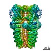 6267MC 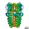 6268C 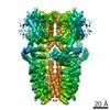 6269C M: map data used to model this data C: citing same article ( |
|---|---|
| Similar structure data | |
| EM raw data | EMPIAR-10024 (Title: Human TRPA1 ion channel with agonist AITC / Data size: 453.0 Data #1: TRP ion channel dataset [picked particles - single frame - processed]) |

-Links
 PDBj
PDBj


- Assembly
| Deposited unit | 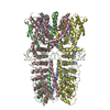
|
|---|---|
| 1 |
|
-Components
| #1: Protein | Mass: 172575.562 Da / Num. of mol.: 4 / Fragment: SEE REMARK 999 Source method: isolated from a genetically manipulated source Source: (gene. exp.) Escherichia coli, Homo sapiens / Gene: malE, ANKTM1, TRPA1 / Cell line (production host): HEK293 GnTi- / Production host: Homo sapiens (human) / References: UniProt: P0AEX9, UniProt: O75762Sequence details | PROTEIN IS A CHIMERA COMPRISING | |
|---|
-Experimental details
-Experiment
| Experiment | Method: ELECTRON MICROSCOPY |
|---|---|
| EM experiment | Aggregation state: PARTICLE / 3D reconstruction method: single particle reconstruction |
- Sample preparation
| Component | Name: Recombinant human TRPA1 ion channel / Type: COMPLEX |
|---|---|
| Buffer solution | Name: 20 mM HEPES, 150 mM NaCl, 1 mM DTT, 1 mM IP6 / pH: 8 / Details: 20 mM HEPES, 150 mM NaCl, 1 mM DTT, 1 mM IP6 |
| Specimen | Conc.: 0.5 mg/ml / Embedding applied: NO / Shadowing applied: NO / Staining applied: NO / Vitrification applied: YES |
| Specimen support | Details: 400 mesh holey carbon |
| Vitrification | Instrument: FEI VITROBOT MARK I / Cryogen name: ETHANE / Temp: 120 K / Humidity: 100 % Details: Blot for 7 seconds before plunging into liquid ethane (FEI VITROBOT MARK I). Method: Blot for 7 seconds before plunging. |
- Electron microscopy imaging
| Experimental equipment |  Model: Tecnai Polara / Image courtesy: FEI Company |
|---|---|
| Microscopy | Model: FEI POLARA 300 / Date: Jul 18, 2014 Details: Gatan K2 Summit in super-resolution counting mode. Motion correction as described in Li et al. (2013) Nature Methods. |
| Electron gun | Electron source:  FIELD EMISSION GUN / Accelerating voltage: 300 kV / Illumination mode: FLOOD BEAM FIELD EMISSION GUN / Accelerating voltage: 300 kV / Illumination mode: FLOOD BEAM |
| Electron lens | Mode: BRIGHT FIELD / Nominal magnification: 31000 X / Calibrated magnification: 31000 X / Nominal defocus max: 2800 nm / Nominal defocus min: 1500 nm / Cs: 2 mm |
| Specimen holder | Temperature: 120 K |
| Image recording | Electron dose: 21 e/Å2 / Film or detector model: GATAN K2 (4k x 4k) Details: Gatan K2 Summit in super-resolution counting mode. Motion correction as described in Li et al. (2013) Nature Methods. |
| Image scans | Num. digital images: 1160 |
| Radiation | Protocol: SINGLE WAVELENGTH / Monochromatic (M) / Laue (L): M / Scattering type: x-ray |
| Radiation wavelength | Relative weight: 1 |
- Processing
| EM software | Name: RELION / Category: 3D reconstruction | ||||||||||||
|---|---|---|---|---|---|---|---|---|---|---|---|---|---|
| CTF correction | Details: Each particle | ||||||||||||
| Symmetry | Point symmetry: C4 (4 fold cyclic) | ||||||||||||
| 3D reconstruction | Method: Maximum likelihood / Resolution: 4.24 Å / Resolution method: FSC 0.143 CUT-OFF / Num. of particles: 43585 / Nominal pixel size: 1.2156 Å / Actual pixel size: 1.2156 Å / Symmetry type: POINT | ||||||||||||
| Refinement step | Cycle: LAST
|
