 Movie
Movie Controller


+ Open data

- Basic information
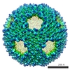
| Entry | Database: PDB / ID: 3j8v | ||||||
|---|---|---|---|---|---|---|---|
| Title | Cryo-EM reconstruction of quasi-HPV16 complex with H16.14J Fab | ||||||
 Components Components |
| ||||||
 Keywords Keywords | VIRUS/IMMUNE SYSTEM / L1 pentamer / quasi-HPV16 / L1 capsomer / Rosie online / VIRUS-IMMUNE SYSTEM complex | ||||||
| Function / homology |  Function and homology information Function and homology informationT=7 icosahedral viral capsid / endocytosis involved in viral entry into host cell / virion attachment to host cell / host cell nucleus / structural molecule activity Similarity search - Function | ||||||
| Biological species |  Human papillomavirus type 16 Human papillomavirus type 16 | ||||||
| Method | ELECTRON MICROSCOPY / single particle reconstruction / cryo EM / Resolution: 13.9 Å | ||||||
 Authors Authors | Guan, J. / Hafenstein, S. | ||||||
 Citation Citation | Journal: Virology / Year: 2015 Title: Structural comparison of four different antibodies interacting with human papillomavirus 16 and mechanisms of neutralization. Authors: Jian Guan / Stephanie M Bywaters / Sarah A Brendle / Hyunwook Lee / Robert E Ashley / Alexander M Makhov / James F Conway / Neil D Christensen / Susan Hafenstein /  Abstract: Cryo-electron microscopy (cryo-EM) was used to solve the structures of human papillomavirus type 16 (HPV16) complexed with fragments of antibody (Fab) from three different neutralizing monoclonals ...Cryo-electron microscopy (cryo-EM) was used to solve the structures of human papillomavirus type 16 (HPV16) complexed with fragments of antibody (Fab) from three different neutralizing monoclonals (mAbs): H16.1A, H16.14J, and H263.A2. The structure-function analysis revealed predominantly monovalent binding of each Fab with capsid interactions that involved multiple loops from symmetry related copies of the major capsid protein. The residues identified in each Fab-virus interface map to a conformational groove on the surface of the capsomer. In addition to the known involvement of the FG and HI loops, the DE loop was also found to constitute the core of each epitope. Surprisingly, the epitope mapping also identified minor contributions by EF and BC loops. Complementary immunological assays included mAb and Fab neutralization. The specific binding characteristics of mAbs correlated with different neutralizing behaviors in pre- and post-attachment neutralization assays. | ||||||
| History |
|
- Structure visualization
| Movie |
Movie viewer |
|---|---|
| Structure viewer | Molecule: MolmilJmol/JSmol |
 UCSF Chimera
- Downloads & links
-Download
| PDBx/mmCIF format | 3j8v.cif.gz | 689.6 KB | Display | PDBx/mmCIF format |
|---|---|---|---|---|
| PDB format | pdb3j8v.ent.gz | 568.5 KB | Display | PDB format |
| PDBx/mmJSON format | 3j8v.json.gz | Tree view | PDBx/mmJSON format | |
| Others |  Other downloads Other downloads |
-Validation report
| Summary document | 3j8v_validation.pdf.gz | 846.7 KB | Display | wwPDB validaton report |
|---|---|---|---|---|
| Full document | 3j8v_full_validation.pdf.gz | 935.1 KB | Display | |
| Data in XML | 3j8v_validation.xml.gz | 88.3 KB | Display | |
| Data in CIF | 3j8v_validation.cif.gz | 130.5 KB | Display | |
| Arichive directory | https://data.pdbj.org/pub/pdb/validation_reports/j8/3j8vftp://data.pdbj.org/pub/pdb/validation_reports/j8/3j8v | HTTPS FTP |
-Related structure data
| Related structure data | 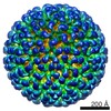 6121MC 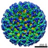 5990C 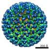 6184C 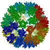 3j8wC  3j8zC M: map data used to model this data C: citing same article ( |
|---|---|
| Similar structure data |


-Links
 PDBj
PDBj


- Assembly
| Deposited unit | 
|
|---|---|
| 1 | x 60
|
| 2 |
|
| 3 | x 5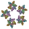
|
| 4 | x 6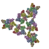
|
| 5 | 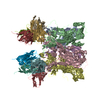
|
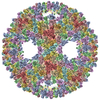
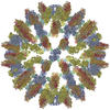
| Symmetry | Point symmetry: (Schoenflies symbol: I (icosahedral)) |
-Components
| #1: Protein | Mass: 50873.422 Da / Num. of mol.: 5 / Fragment: UNP residues 47-500 / Source method: isolated from a natural source / Source: (natural) Human papillomavirus type 16 / References: UniProt: Q4VRM0, UniProt: P03101*PLUS#2: Antibody | Mass: 11810.232 Da / Num. of mol.: 4 / Fragment: variable domain Fab / Source method: isolated from a natural source / Source: (natural) #3: Antibody | Mass: 12994.408 Da / Num. of mol.: 4 / Fragment: variable domain Fab / Source method: isolated from a natural source / Source: (natural) Has protein modification | Y | |
|---|
-Experimental details
-Experiment
| Experiment | Method: ELECTRON MICROSCOPY |
|---|---|
| EM experiment | Aggregation state: PARTICLE / 3D reconstruction method: single particle reconstruction |
- Sample preparation
| Component |
| ||||||||||||||||||||
|---|---|---|---|---|---|---|---|---|---|---|---|---|---|---|---|---|---|---|---|---|---|
| Molecular weight | Value: 44.7 MDa / Experimental value: NO | ||||||||||||||||||||
| Details of virus | Empty: NO / Enveloped: NO / Host category: VERTEBRATES / Isolate: OTHER / Type: VIRION | ||||||||||||||||||||
| Natural host | Organism: Homo sapiens | ||||||||||||||||||||
| Buffer solution | Name: 1 M NaCl, 200 mM Tris / pH: 7.4 / Details: 1 M NaCl, 200 mM Tris | ||||||||||||||||||||
| Specimen | Conc.: 1 mg/ml / Embedding applied: NO / Shadowing applied: NO / Staining applied: NO / Vitrification applied: YES | ||||||||||||||||||||
| Specimen support | Details: glow-discharged holey carbon support grid | ||||||||||||||||||||
| Vitrification | Instrument: GATAN CRYOPLUNGE 3 / Cryogen name: ETHANE / Temp: 102 K / Humidity: 90 % Details: Blot for 0.7 seconds before plunging into liquid ethane (GATAN CRYOPLUNGE 3) Method: Blot for 0.7 seconds before plunging. |
- Electron microscopy imaging
| Microscopy | Model: JEOL 2100 / Date: Aug 27, 2014 |
|---|---|
| Electron gun | Electron source: LAB6 / Accelerating voltage: 200 kV / Illumination mode: SPOT SCAN |
| Electron lens | Mode: BRIGHT FIELD / Nominal magnification: 50000 X / Nominal defocus max: 4690 nm / Nominal defocus min: 620 nm / Cs: 2 mm / Camera length: 0 mm |
| Specimen holder | Specimen holder model: GATAN LIQUID NITROGEN / Temperature: 95 K |
| Image recording | Electron dose: 15 e/Å2 / Film or detector model: GATAN ULTRASCAN 4000 (4k x 4k) |
| Image scans | Num. digital images: 385 |
| Radiation | Protocol: SINGLE WAVELENGTH / Monochromatic (M) / Laue (L): M / Scattering type: x-ray |
| Radiation wavelength | Relative weight: 1 |
- Processing
| EM software |
| ||||||||||||||||||||
|---|---|---|---|---|---|---|---|---|---|---|---|---|---|---|---|---|---|---|---|---|---|
| CTF correction | Details: Each particle | ||||||||||||||||||||
| Symmetry | Point symmetry: I (icosahedral) | ||||||||||||||||||||
| 3D reconstruction | Method: Cross-common Lines / Resolution: 13.9 Å / Resolution method: FSC 0.5 CUT-OFF / Num. of particles: 5642 / Nominal pixel size: 2.33 Å / Actual pixel size: 2.33 Å Details: Semi-automatic particle selection was performed using e2boxer.py to obtain the particle coordinates, followed by particle boxing, linearization, normalization, and apodization of the images ...Details: Semi-automatic particle selection was performed using e2boxer.py to obtain the particle coordinates, followed by particle boxing, linearization, normalization, and apodization of the images using Robem. Defocus and astigmatism values necessary to perform contrast transfer function (CTF) correction for the extracted particles were assessed using Robem. The icosahedrally averaged reconstruction was initiated using a random model generated with setup_rmc. For the last step of refinement, the final maps were CTF-corrected using a B factor of 200 A2. (Single particle--Applied symmetry: I) Symmetry type: POINT | ||||||||||||||||||||
| Atomic model building | Protocol: RIGID BODY FIT / Space: REAL / Details: REFINEMENT PROTOCOL--rigid body | ||||||||||||||||||||
| Atomic model building | 3D fitting-ID: 1 / Accession code: 3OAE / Initial refinement model-ID: 1 / PDB-ID: 3OAE  3oae
| ||||||||||||||||||||
| Refinement step | Cycle: LAST
|
