 Movie
Movie Controller

[English] 日本語
 Yorodumi
- PDB-3j8h: Structure of the rabbit ryanodine receptor RyR1 in complex with F... -
+ Open data

- Basic information
| Entry | Database: PDB / ID: 3j8h | ||||||
|---|---|---|---|---|---|---|---|
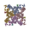
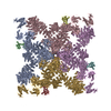
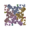
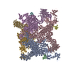
| Title | Structure of the rabbit ryanodine receptor RyR1 in complex with FKBP12 at 3.8 Angstrom resolution | ||||||
 Components Components |
| ||||||
 Keywords Keywords | TRANSPORT PROTEIN/ISOMERASE / rabbit ryanodine receptor RyR1 / high-conductance intracellular Ca2+ channels / excitation-contraction coupling / TRANSPORT PROTEIN-ISOMERASE complex | ||||||
| Function / homology |  Function and homology information Function and homology informationcytoplasmic side of membrane / ATP-gated ion channel activity / terminal cisterna / ryanodine receptor complex / ryanodine-sensitive calcium-release channel activity / release of sequestered calcium ion into cytosol by sarcoplasmic reticulum / ossification involved in bone maturation / cellular response to caffeine / skin development / organelle membrane ...cytoplasmic side of membrane / ATP-gated ion channel activity / terminal cisterna / ryanodine receptor complex / ryanodine-sensitive calcium-release channel activity / release of sequestered calcium ion into cytosol by sarcoplasmic reticulum / ossification involved in bone maturation / cellular response to caffeine / skin development / organelle membrane / intracellularly gated calcium channel activity / smooth endoplasmic reticulum / outflow tract morphogenesis / regulation of ryanodine-sensitive calcium-release channel activity / toxic substance binding / striated muscle contraction / voltage-gated calcium channel activity / skeletal muscle fiber development / regulation of release of sequestered calcium ion into cytosol by sarcoplasmic reticulum / release of sequestered calcium ion into cytosol / sarcoplasmic reticulum membrane / muscle contraction / cellular response to calcium ion / sarcoplasmic reticulum / peptidylprolyl isomerase / peptidyl-prolyl cis-trans isomerase activity / sarcolemma / calcium ion transmembrane transport / calcium channel activity / Z disc / intracellular calcium ion homeostasis / disordered domain specific binding / protein homotetramerization / transmembrane transporter binding / calmodulin binding / intracellular membrane-bounded organelle / calcium ion binding / ATP binding / identical protein binding / membrane / cytosol Similarity search - Function | ||||||
| Biological species |  | ||||||
| Method | ELECTRON MICROSCOPY / single particle reconstruction / cryo EM / Resolution: 3.8 Å | ||||||
 Authors Authors | Yan, Z. / Bai, X. / Yan, C. / Wu, J. / Scheres, S.H.W. / Shi, Y. / Yan, N. | ||||||
 Citation Citation | Journal: Nature / Year: 2015 Title: Structure of the rabbit ryanodine receptor RyR1 at near-atomic resolution. Authors: Zhen Yan / Xiaochen Bai / Chuangye Yan / Jianping Wu / Zhangqiang Li / Tian Xie / Wei Peng / Changcheng Yin / Xueming Li / Sjors H W Scheres / Yigong Shi / Nieng Yan /   Abstract: The ryanodine receptors (RyRs) are high-conductance intracellular Ca(2+) channels that play a pivotal role in the excitation-contraction coupling of skeletal and cardiac muscles. RyRs are the largest ...The ryanodine receptors (RyRs) are high-conductance intracellular Ca(2+) channels that play a pivotal role in the excitation-contraction coupling of skeletal and cardiac muscles. RyRs are the largest known ion channels, with a homotetrameric organization and approximately 5,000 residues in each protomer. Here we report the structure of the rabbit RyR1 in complex with its modulator FKBP12 at an overall resolution of 3.8 Å, determined by single-particle electron cryomicroscopy. Three previously uncharacterized domains, named central, handle and helical domains, display the armadillo repeat fold. These domains, together with the amino-terminal domain, constitute a network of superhelical scaffold for binding and propagation of conformational changes. The channel domain exhibits the voltage-gated ion channel superfamily fold with distinct features. A negative-charge-enriched hairpin loop connecting S5 and the pore helix is positioned above the entrance to the selectivity-filter vestibule. The four elongated S6 segments form a right-handed helical bundle that closes the pore at the cytoplasmic border of the membrane. Allosteric regulation of the pore by the cytoplasmic domains is mediated through extensive interactions between the central domains and the channel domain. These structural features explain high ion conductance by RyRs and the long-range allosteric regulation of channel activities. | ||||||
| History |
|
- Structure visualization
| Movie |
Movie viewer |
|---|---|
| Structure viewer | Molecule: MolmilJmol/JSmol |
- Downloads & links
-Download
| PDBx/mmCIF format | 3j8h.cif.gz | 2.5 MB | Display | PDBx/mmCIF format |
|---|---|---|---|---|
| PDB format | pdb3j8h.ent.gz | Display | PDB format | |
| PDBx/mmJSON format | 3j8h.json.gz | Tree view | PDBx/mmJSON format | |
| Others |  Other downloads Other downloads |
-Validation report
| Summary document | 3j8h_validation.pdf.gz | 1.2 MB | Display | wwPDB validaton report |
|---|---|---|---|---|
| Full document | 3j8h_full_validation.pdf.gz | 1.7 MB | Display | |
| Data in XML | 3j8h_validation.xml.gz | 399.8 KB | Display | |
| Data in CIF | 3j8h_validation.cif.gz | 611 KB | Display | |
| Arichive directory | https://data.pdbj.org/pub/pdb/validation_reports/j8/3j8hftp://data.pdbj.org/pub/pdb/validation_reports/j8/3j8h | HTTPS FTP |
-Related structure data
| Related structure data | 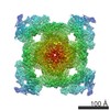 2807MC M: map data used to model this data C: citing same article ( |
|---|---|
| Similar structure data |


-Links
 PDBj
PDBj

- Assembly
| Deposited unit | 
|
|---|---|
| 1 |
|
-Components
| #1: Protein | Mass: 500211.562 Da / Num. of mol.: 4 / Source method: isolated from a natural source / Source: (natural) #2: Protein | Mass: 11836.508 Da / Num. of mol.: 4 Source method: isolated from a genetically manipulated source Source: (gene. exp.)  #3: Chemical | ChemComp-ZN /   Mass: 65.409 Da / Num. of mol.: 4 / Source method: obtained synthetically / Formula: Zn Mass: 65.409 Da / Num. of mol.: 4 / Source method: obtained synthetically / Formula: Zn |
|---|
-Experimental details
-Experiment
| Experiment | Method: ELECTRON MICROSCOPY |
|---|---|
| EM experiment | Aggregation state: PARTICLE / 3D reconstruction method: single particle reconstruction |
- Sample preparation
| Component |
| ||||||||||||||||
|---|---|---|---|---|---|---|---|---|---|---|---|---|---|---|---|---|---|
| Buffer solution | Name: 20 mM MOPS sodium, pH 7.4, 250 mM NaCl, 2 mM DTT, 2 mM EGTA, 0.015% Tween20, protein inhibitors pH: 7.4 Details: 20 mM MOPS sodium, pH 7.4, 250 mM NaCl, 2 mM DTT, 2 mM EGTA, 0.015% Tween20, protein inhibitors | ||||||||||||||||
| Specimen | Conc.: 0.06 mg/ml / Embedding applied: NO / Shadowing applied: NO / Staining applied: NO / Vitrification applied: YES | ||||||||||||||||
| Specimen support | Details: holey carbon grids (Quantifoil CuR2/2), on which a home-made continuous carbon film (estimated to be ~30 Angstrom thick) had previously been deposited | ||||||||||||||||
| Vitrification | Instrument: FEI VITROBOT MARK III / Cryogen name: NITROGEN / Humidity: 100 % Details: Protein solution was applied to glow-discharged carbon coated grid and blotted for 2 seconds before plunging into liquid nitrogen (FEI VITROBOT MARK III) Method: protein solution was applied to glow-discharged carbon coated grid and blotted for 2 seconds before plunging |
- Electron microscopy imaging
| Experimental equipment |  Model: Tecnai Polara / Image courtesy: FEI Company |
|---|---|
| Microscopy | Model: FEI POLARA 300 / Date: Jun 21, 2014 |
| Electron gun | Electron source:  FIELD EMISSION GUN / Accelerating voltage: 300 kV / Illumination mode: FLOOD BEAM FIELD EMISSION GUN / Accelerating voltage: 300 kV / Illumination mode: FLOOD BEAM |
| Electron lens | Mode: BRIGHT FIELD / Nominal magnification: 78000 X / Calibrated magnification: 104748 X / Nominal defocus max: 5600 nm / Nominal defocus min: 1900 nm / Cs: 2 mm |
| Specimen holder | Specimen holder type: GATAN LIQUID NITROGEN / Temperature: 85 K / Temperature (max): 90 K / Temperature (min): 80 K |
| Image recording | Electron dose: 40 e/Å2 / Film or detector model: FEI FALCON II (4k x 4k) |
| Radiation wavelength | Relative weight: 1 |
- Processing
| Software | Name: REFMAC / Version: 5.8.0049 2013/08/13 / Classification: refinement / Contact author: Garib N. Murshudov / Contact author email: garib[at]mrc-lmb.cam.ac.uk Description: (un)restrained refinement or idealisation of macromolecular structures | ||||||||||||||||||||||||
|---|---|---|---|---|---|---|---|---|---|---|---|---|---|---|---|---|---|---|---|---|---|---|---|---|---|
| EM software |
| ||||||||||||||||||||||||
| CTF correction | Details: Contrast transfer function parameters were estimated using CTFFIND3 | ||||||||||||||||||||||||
| Symmetry | Point symmetry: C4 (4 fold cyclic) | ||||||||||||||||||||||||
| 3D reconstruction | Resolution: 3.8 Å / Resolution method: FSC 0.143 CUT-OFF / Num. of particles: 65872 / Nominal pixel size: 1.34 Å / Actual pixel size: 1.34 Å Details: All two- and three-dimensional classifications and refinements were performed using RELION 1.3. The model was built manually in COOT, and was refined in real space using Phenix and in ...Details: All two- and three-dimensional classifications and refinements were performed using RELION 1.3. The model was built manually in COOT, and was refined in real space using Phenix and in reciprocal space using Refmac. Symmetry type: POINT | ||||||||||||||||||||||||
| Refinement | Details: HYDROGENS HAVE BEEN ADDED IN THEIR RIDING POSITIONS | ||||||||||||||||||||||||
| Refinement step | Cycle: LAST
|
