 Movie
Movie Controller


+ Open data

- Basic information
| Entry | Database: PDB / ID: 3j5l | ||||||
|---|---|---|---|---|---|---|---|
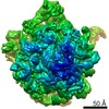
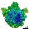
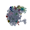
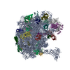
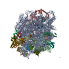
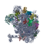
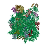
| Title | Structure of the E. coli 50S subunit with ErmBL nascent chain | ||||||
 Components Components |
| ||||||
 Keywords Keywords | RIBOSOME/ANTIBIOTIC / erythromycin / stalling / RIBOSOME-ANTIBIOTIC complex | ||||||
| Function / homology |  Function and homology information Function and homology informationstringent response / translational termination / transcriptional attenuation / endoribonuclease inhibitor activity / positive regulation of ribosome biogenesis / RNA-binding transcription regulator activity / negative regulation of cytoplasmic translation / DnaA-L2 complex / translation repressor activity / negative regulation of DNA-templated DNA replication initiation ...stringent response / translational termination / transcriptional attenuation / endoribonuclease inhibitor activity / positive regulation of ribosome biogenesis / RNA-binding transcription regulator activity / negative regulation of cytoplasmic translation / DnaA-L2 complex / translation repressor activity / negative regulation of DNA-templated DNA replication initiation / mRNA regulatory element binding translation repressor activity / response to reactive oxygen species / cytosolic ribosome assembly / ribosome assembly / assembly of large subunit precursor of preribosome / regulation of cell growth / DNA-templated transcription termination / response to radiation / mRNA 5'-UTR binding / large ribosomal subunit / transferase activity / ribosome binding / 5S rRNA binding / ribosomal large subunit assembly / large ribosomal subunit rRNA binding / cytosolic large ribosomal subunit / cytoplasmic translation / tRNA binding / negative regulation of translation / rRNA binding / structural constituent of ribosome / ribosome / translation / response to antibiotic / negative regulation of DNA-templated transcription / mRNA binding / DNA binding / RNA binding / zinc ion binding / cytoplasm / cytosol Similarity search - Function | ||||||
| Biological species |  Streptococcus sanguinis (bacteria) Streptococcus sanguinis (bacteria) | ||||||
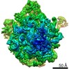
| Method | ELECTRON MICROSCOPY / single particle reconstruction / cryo EM / Resolution: 6.6 Å | ||||||
 Authors Authors | Arenz, S. / Ramu, H. / Gupta, P. / Berninghausen, O. / Beckmann, R. / Vazquez-Laslop, N. / Mankin, A.S. / Wilson, D.N. | ||||||
 Citation Citation | Journal: Nat Commun / Year: 2014 Title: Molecular basis for erythromycin-dependent ribosome stalling during translation of the ErmBL leader peptide. Authors: Stefan Arenz / Haripriya Ramu / Pulkit Gupta / Otto Berninghausen / Roland Beckmann / Nora Vázquez-Laslop / Alexander S Mankin / Daniel N Wilson /   Abstract: In bacteria, ribosome stalling during translation of ErmBL leader peptide occurs in the presence of the antibiotic erythromycin and leads to induction of expression of the downstream macrolide ...In bacteria, ribosome stalling during translation of ErmBL leader peptide occurs in the presence of the antibiotic erythromycin and leads to induction of expression of the downstream macrolide resistance methyltransferase ErmB. The lack of structures of drug-dependent stalled ribosome complexes (SRCs) has limited our mechanistic understanding of this regulatory process. Here we present a cryo-electron microscopy structure of the erythromycin-dependent ErmBL-SRC. The structure reveals that the antibiotic does not interact directly with ErmBL, but rather redirects the path of the peptide within the tunnel. Furthermore, we identify a key peptide-ribosome interaction that defines an important relay pathway from the ribosomal tunnel to the peptidyltransferase centre (PTC). The PTC of the ErmBL-SRC appears to adopt an uninduced state that prevents accommodation of Lys-tRNA at the A-site, thus providing structural basis for understanding how the drug and the nascent peptide cooperate to inhibit peptide bond formation and induce translation arrest. | ||||||
| History |
|
- Structure visualization
| Movie |
Movie viewer |
|---|---|
| Structure viewer | Molecule: MolmilJmol/JSmol |
- Downloads & links
-Download
| PDBx/mmCIF format | 3j5l.cif.gz | 2.2 MB | Display | PDBx/mmCIF format |
|---|---|---|---|---|
| PDB format | pdb3j5l.ent.gz | 1.7 MB | Display | PDB format |
| PDBx/mmJSON format | 3j5l.json.gz | Tree view | PDBx/mmJSON format | |
| Others |  Other downloads Other downloads |
-Validation report
| Arichive directory | https://data.pdbj.org/pub/pdb/validation_reports/j5/3j5lftp://data.pdbj.org/pub/pdb/validation_reports/j5/3j5l | HTTPS FTP |
|---|
-Related structure data
| Related structure data | 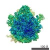 5771MC M: map data used to model this data C: citing same article ( |
|---|---|
| Similar structure data |
-Links
 PDBj
PDBj


- Assembly
| Deposited unit | 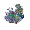
|
|---|---|
| 1 |
|
-Components
+50S ribosomal protein ... , 29 types, 29 molecules 01234CDEFGHIJKLMNOPQRSTUVWXYZ
-RNA chain , 4 types, 4 molecules 57AB
| #6: RNA chain | Mass: 617.483 Da / Num. of mol.: 1 / Source method: obtained synthetically / Details: 3'-end of A-site tRNA |
|---|---|
| #8: RNA chain | Mass: 894.612 Da / Num. of mol.: 1 / Source method: obtained synthetically / Details: 3'-end of P-site tRNA |
| #9: RNA chain | Mass: 941612.375 Da / Num. of mol.: 1 / Source method: isolated from a natural source / Source: (natural) |
| #10: RNA chain | Mass: 38177.762 Da / Num. of mol.: 1 / Source method: isolated from a natural source / Source: (natural) |
-Protein/peptide , 1 types, 1 molecules 6
| #7: Protein/peptide | Mass: 1253.513 Da / Num. of mol.: 1 / Fragment: nascent chain / Source method: isolated from a natural source / Source: (natural) Streptococcus sanguinis (bacteria) |
|---|
-Non-polymers , 2 types, 2 molecules 
| #35: Chemical | ChemComp-UNL / Num. of mol.: 1 / Source method: obtained synthetically |
|---|---|
| #36: Chemical | ChemComp-ERY /  Mass: 733.927 Da / Num. of mol.: 1 / Source method: obtained synthetically / Formula: C37H67NO13 / Comment: antibiotic*YM Mass: 733.927 Da / Num. of mol.: 1 / Source method: obtained synthetically / Formula: C37H67NO13 / Comment: antibiotic*YM |
-Details
| Has protein modification | Y |
|---|---|
| Sequence details | FULL-LENGTH PROTEIN L9 IS PRESENT IN THE RIBOSOME, BUT ONLY RESIDUES 1-56 ARE MODELED. |
-Experimental details
-Experiment
| Experiment | Method: ELECTRON MICROSCOPY |
|---|---|
| EM experiment | Aggregation state: PARTICLE / 3D reconstruction method: single particle reconstruction |
- Sample preparation
| Component | Name: ErmBL-stalled E. coli 70S ribosome / Type: RIBOSOME |
|---|---|
| Buffer solution | pH: 7.4 |
| Specimen | Embedding applied: NO / Shadowing applied: NO / Staining applied: NO / Vitrification applied: YES |
| Vitrification | Instrument: FEI VITROBOT MARK IV / Cryogen name: ETHANE / Details: Plunged into liquid ethane (FEI VITROBOT MARK IV) |
- Electron microscopy imaging
| Experimental equipment |  Model: Titan Krios / Image courtesy: FEI Company |
|---|---|
| Microscopy | Model: FEI TITAN KRIOS / Date: Mar 14, 2013 |
| Electron gun | Electron source:  FIELD EMISSION GUN / Accelerating voltage: 200 kV / Illumination mode: SPOT SCAN FIELD EMISSION GUN / Accelerating voltage: 200 kV / Illumination mode: SPOT SCAN |
| Electron lens | Mode: BRIGHT FIELD / Nominal magnification: 148721 X / Calibrated magnification: 148721 X / Nominal defocus max: 4500 nm / Nominal defocus min: 1000 nm / Cs: 2.7 mm / Camera length: 0 mm |
| Specimen holder | Specimen holder model: FEI TITAN KRIOS AUTOGRID HOLDER / Tilt angle max: 0 ° / Tilt angle min: 0 ° |
| Image recording | Electron dose: 20 e/Å2 / Film or detector model: TVIPS TEMCAM-F416 (4k x 4k) |
| Image scans | Num. digital images: 17906 |
| Radiation | Protocol: SINGLE WAVELENGTH / Monochromatic (M) / Laue (L): M / Scattering type: x-ray |
| Radiation wavelength | Relative weight: 1 |
- Processing
| EM software |
| ||||||||||||
|---|---|---|---|---|---|---|---|---|---|---|---|---|---|
| CTF correction | Details: Defocus groups | ||||||||||||
| Symmetry | Point symmetry: C1 (asymmetric) | ||||||||||||
| 3D reconstruction | Resolution: 6.6 Å / Resolution method: FSC 0.5 CUT-OFF / Num. of particles: 349744 / Nominal pixel size: 1.0605 Å / Actual pixel size: 1.0605 Å / Details: (Single particle--Applied symmetry: C1) / Symmetry type: POINT | ||||||||||||
| Atomic model building | Protocol: RIGID BODY FIT / Space: REAL / Details: REFINEMENT PROTOCOL--rigid body | ||||||||||||
| Atomic model building | PDB-ID: 3OFR 3ofr Accession code: 3OFR / Source name: PDB / Type: experimental model | ||||||||||||
| Refinement step | Cycle: LAST
|
